
Published on
Download the article PDF: Clinical Image Challenges September 2026
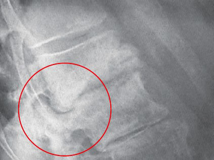
Differential Diagnosis
- Acute discogenic pain
- Vertebral compression fracture
- Soft tissue back injury
- Cauda equina syndrome
- Spondylolisthesis exacerbation
Diagnosis
The radiographic findings include an anterior wedge compression deformity of L1 with loss of anterior vertebral body height, consistent with a vertebral compression fracture. It also shows diffuse osteopenia and intense sclerosis with irregularity of the inferior endplate of L1, which raises concern for pathologic fracture. Multilevel degenerative changes are also visible including disc space narrowing, anterior osteophyte formation, and possible grade I degenerative spondylolisthesis at L4-L5.
Osteoporotic fractures are those occurring from a low-impact injury, such as a fall from a standing height or less. Vertebral compression fractures are the most common type of osteoporotic fracture and most often occur at the midthoracic (T7-T8) spine and the thoracolumbar junction (T12-L1). These types of fractures can cause significant back pain, limit physical functioning and activities of daily living, and lead to loss of independence, depression, and chronic pain. A history of an osteoporotic fracture is the most important risk factor for subsequent fractures; approximately 19 % of patients who have a vertebral compression fracture will have another fracture in the next year.
What to Look For
- Neurologic abnormalities (such as dermatomal sensory deficits, focal weakness or clonus) may indicate the presence of bone fragments in the spinal canal or foramina, which may require urgent surgical intervention.
- Ask about changes in height.Each vertebral compression fracture causes approximately ≥1 cm loss in height; kyphosis may be an indicator of multiple past vertebral compression fractures.
- In any patient presenting with fever or other signs of infection, a complete blood count and blood cultures should be obtained immediately.
Pearls For Urgent Care Management
- In patients with severe pain and/or suspected fracture-related complications, prompt referral to the emergency department for magnetic resonance imaging is indicated.
- Initial management of osteoporotic vertebral compression fractures should include pain control and activity modification. Patients with mild to moderate pain can be treated initially with nonopioid oral analgesics, considering risks of nonsteroidal anti-inflammatory drugs use in older patients.
- Intranasal calcitonin has been shown to hasten the relief of pain from osteoporotic vertebral fracture when initiated early.
- Patients with a tolerable pain level should be encouraged to resume physical activity as quickly as possible. Complete bed rest is not recommended and can lead to further bone loss and deconditioning. Physical therapy and aquatic therapy are recommended for strengthening and pain management.
- Patient Education: fractures may take up to 3 months to heal, and that pain and activity intolerance should diminish gradually.
- Bracing and muscle relaxants are not recommended.
- Encourage follow-up with primary care provider for long-term evaluation and management of osteoporosis.
- Consider referral to orthopedic and/or spine specialist, as patients who continue to have refractory pain may sometimes be candidates for surgical interventions for vertebral fractures.
Read More
- Back Pain From Vertebral Metastases in Prostate Cancer: A Case Report
- Abdominal Pseudohernia and Urinary Retention Due to Spinal Nerve Root Compression From Disc Herniation: A Case Report
- Urgent Care Management of Geriatric Falls
- Back Pain, an Urgent Care Visit—and a Devastating Outcome
- A 35-Year-Old with Neck Pain After an Auto Accident
Images provided by Experity Teleradiology