
Published on
Download the article PDF: Urgent Care Diagnosis And Management Of Toddlers Fracture
Urgent Message: Identifying fall height and mechanism, while also relying on caregiver narratives, can help urgent care clinicians rule out severe pathologies and diagnose a toddler’s fracture in ambulatory pediatric patients after a fall.
Connor Osmond, BA; Thomas Seebacher, DO
Keywords: fractures, bone; tibial fractures; gait disorders, neurologic; radiography; accidental falls
Editor’s Note: While the images presented here are authentic, the patient case scenario is hypothetical.
Questions for the Clinician at the Bedside
- Which mechanisms and patient features are associated with a toddler’s fracture?
- What are the red flag signs that should be considered in patients with a suspected toddler’s fracture?
- What radiographic findings are suggestive of a toddler’s fracture, and why are x-rays often negative?
- Which patients require immobilization vs immediate emergency department referral?
- What are the complications of missing the diagnosis of a toddler’s fracture?
Abstract
A toddler’s fracture is a nondisplaced spiral fracture of the distal tibia known to occur in pediatric patients between the ages of 9 months and 3 years old. Toddler’s fractures are typically seen in ambulatory pediatric patients showing a sudden refusal to bear weight or presenting with an acute onset of limp after an unwitnessed fall. These fractures result from low-energy pivoting, twisting, or rotational injury when the foot is planted, causing a spiral fracture along the distal tibia. Fibular involvement is uncommon in toddler’s fractures and generally indicates a different fracture pattern. A toddler’s fracture commonly presents with normal radiographs at the initial visit; however, the physical exam will show tenderness with palpation and painful passive dorsiflexion of the ankle.
Clinical Scenario
A previously healthy 20-month-old boy was brought to the urgent care (UC) center by his parents after refusing to bear weight on his right leg since earlier that afternoon. His mother reported that he was running on the carpeted living room floor when he tripped and fell. The mother did not witness the fall directly, but her son reportedly began crying immediately. He had been crawling—rather than walking—since the incident. He had no fever, no recent illness, and no other injuries.
On arrival at UC, the child was in no acute distress and sat comfortably in his mother’s lap. He refused to bear weight on his right leg even with support. Vital signs and growth chart were within normal limits for the child’s age. The right lower extremity revealed no deformity, swelling, or bruising; however, the patient had focal tenderness over the distal one-third of the right tibia upon palpation and pain upon passive dorsiflexion of the ankle.
There was full passive range of motion at the hip and knee, and the neurovascular exam was normal including normal capillary refill in the toes. The x-ray performed in UC appeared normal. The patient was discharged with a controlled ankle motion (CAM) boot and follow-up was scheduled in 3 weeks.
Epidemiology
Toddler’s fracture, also called childhood accidental spiral tibial (CAST) fracture, is a fracture unique to ambulatory infants and young children.[1] These injuries are most commonly seen in children with a median age of 22 months, typically presenting during the peak age range of 1–3 years as they master independent walking.[2] Although they represent a significant portion of early childhood limping presentations, up to 40% of these cases are initially radiographically occult, meaning no clear fracture line is visible during the first emergency department visit.[3] Radiographically occult toddler’s fractures often become more visible on repeat radiographs obtained 1–2 weeks after the initial injury.[4] Thus, misdiagnosis is common during the initial exam, which can result in incorrectly attributing the mechanism to nonaccidental trauma (NAT) or systemic problems.
Understanding the Anatomy
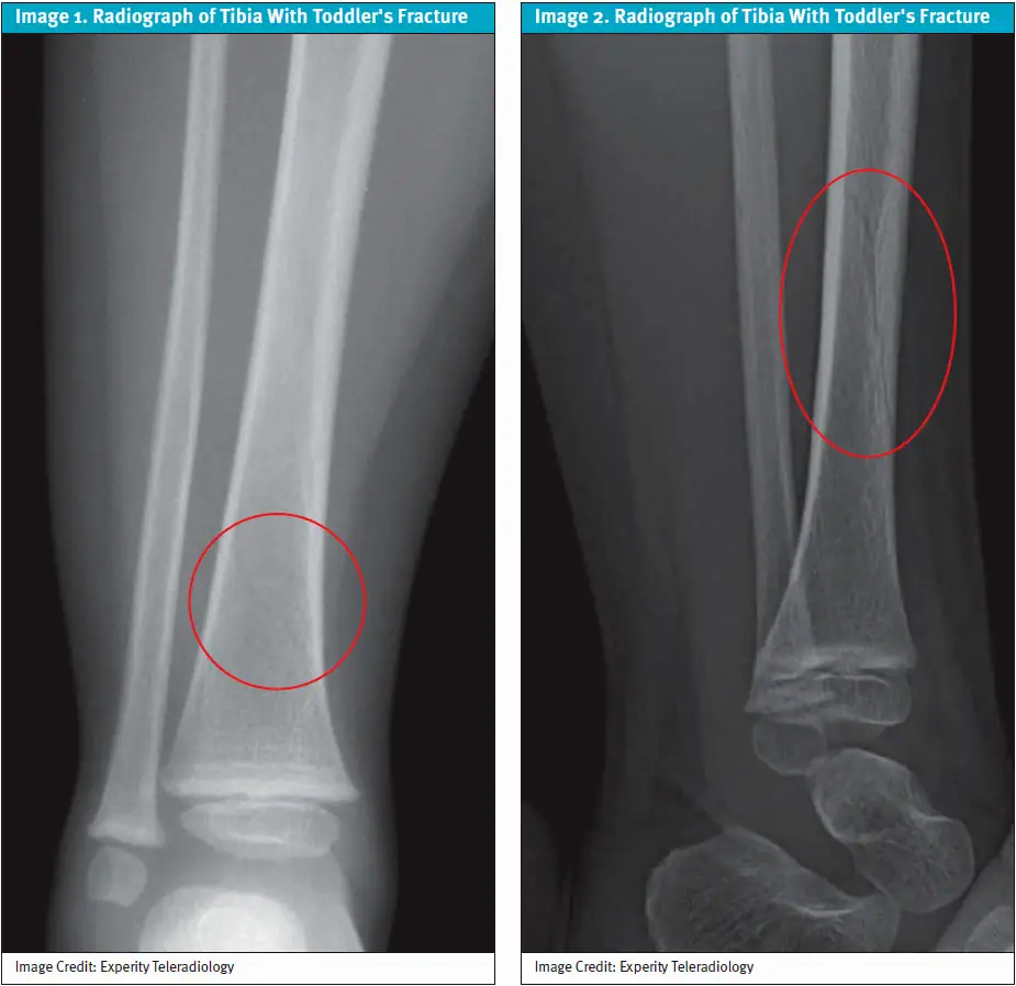
Flexibility, bone density, and growth plate fusion can all weigh into the physician’s consideration for diagnoses of acute fractures, sprains, strains, or NAT. In toddler’s fractures, the primary bone affected is the tibia, typically presenting as a spiral or oblique fracture in the diaphyseal region and often cortical rather than extending into the growth plate (Images 1–2). The fibula, the bone located lateral to the tibia, is rarely involved and usually indicates more significant trauma that would be identified as a different fracture type.[5]
The physis, a cartilaginous structure located between the metaphysis and epiphysis, is often spared as well. The presence of open growth plates helps reflect the overall immature skeletal structure, a key component depicting the nature of these fractures.1
Toddler bones contain a higher proportion of collagen compared to adult bones, making them more flexible and able to absorb more energy without cortical disruption, but they have lower mineral density than mature bone, reducing the resistance against torsional loading.[6]
The thicker periosteum in toddlers provides additional stabilization and limits displacement, which is why toddler’s fractures may be radiographically occult on initial imaging. A periosteal reaction refers to a bone formation stimulated by injury to the periosteum.1,3 In toddler’s fractures, a periosteal reaction may not be visible until 7–14 days after injury, thus causing difficulty in initial diagnosis.
History
Toddler’s fractures most often result from low-energy pivoting or rotational mechanisms—such as tripping, stumbling, or twisting—with minimal physical exam distinctions.1,[7] Caregivers often describe a trivial fall, frequently without visible swelling or deformity, with a presentation of the child’s sudden refusal to bear weight or acute limping. Falls are typically from ground level or less than 1 meter, and caregivers frequently describe the event as minor or insignificant.1,3,[8]
Systemic symptoms such as fever, weight loss, or severe pain are usually absent. Their presence raises concern for more serious conditions like osteomyelitis, septic arthritis, or malignancy, which may also cause refusal to bear weight but involve systemic signs and progressive disease.8,[9],[10] Therefore, fall height, mechanism, and caregiver narratives are essential inputs to help rule out severe pathologies or disease and determine a true toddler’s fracture. Although spiral fractures in ambulatory toddlers are generally benign, similar patterns in nonambulatory children or in the presence of unclear caregiver narratives may warrant further investigation for NAT, such as child abuse.[11]
Physical Examination
UC clinicians should observe the pain threshold and perform a complete physical examination including a gait evaluation. General assessment includes examination of both lower extremities, pelvis, and hips, as toddler’s fractures often present with nonspecific findings.3,8 Evaluation of the affected extremity will show localized point tenderness over the distal one-third of the tibial shaft, minimal or absent swelling, and lack of ecchymosis or deformity.[12] A toddler’s fracture will often present with an antalgic gait, toe-walking, complete refusal to bear weight, or increased irritability with handling of the affected limb.[13] Even if the patient can ambulate, pain or tenderness along the shin can indicate a fracture.[14]
Excluding alternative diagnoses will be one of the most important parts of the physical exam. An ankle sprain or foot contusion may present with localized tenderness and swelling along the foot or ankle but lacks pain with tibial rotation.8 Referred pain from the hip, from diagnoses including osteomyelitis, transient synovitis, and septic arthritis, should always be considered in an injured pediatric patient with gait abnormalities or refusal to bear weight.9 When there is suspected infection, fever, or severe pain of the lower extremity, urgent evaluation is necessary; however, these features often rule out a toddler’s fracture.
Diagnostic Testing
Plain radiographs should include anteroposterior (AP) and lateral tibial views.3 Note that there is a high false-negative rate of around 39% on initial imaging, thus the fracture may be radiographically occult at initial presentation.4,[15]
In a suspected toddler’s fracture, additional imaging rarely changes management. Therefore, escalation to advanced imaging, such as computed tomography (CT), is rarely indicated and exposes the child to unnecessary radiation.2 When radiographs are negative for a tibial spiral fracture, the next priority for management is to rely on physical exam and plan for outpatient follow-up.[16]
Delayed radiographic diagnosis is not known to worsen outcomes for toddler’s fractures and does not alter clinical recovery. Therefore, the patient’s caregivers may need reassurance if diagnosis is achieved on repeat radiograph imaging or outpatient follow-up. Follow-up imaging may demonstrate periosteal reaction or callus formation along the tibial cortex.2,[17]
Differential Diagnosis
A child presenting with a limp may have a wide spectrum of causes, from benign injuries to serious conditions.5 To develop a differential diagnosis, clinical examination should include examination of the legs for bruising, deformity, scars, and skin discoloration as well as a gait evaluation. Additionally, neurologic, abdominal, and lymphadenopathy examinations should be performed as part of the physical assessment to identify other pathologies.[18]
The differential diagnoses include:
- Normal toddler refusal to walk after a minor fall
- Tibial contusion
- Ankle sprain (less common in toddlers)
- Occult foot fracture
- Septic arthritis (hip/knee)
- Transient synovitis (hip)
- Osteomyelitis
- NAT (child abuse)
Indications for Emergency Department Referral
Septic arthritis and osteomyelitis in pediatric patients represent true emergencies and often present subtly with vague pain or refusal to bear weight. These concerns should prompt emergency department (ED) referral.[19]
Additionally, refer the patient immediately with concern for:
- Inability to bear weight and suspicion of systemic infection (eg, fever, toxic appearance)
- Neurovascular compromise (ie, diminished distal pulses)
- Concern for NAT
- Multiple injuries or uncertain mechanism
- Severe swelling suggesting compartment syndrome (rare)
Urgent Care Management
Immobilization and Pain Management
Preferred initial management is immobilization in a CAM boot with referral to a pediatric orthopedist.[20] The boot can be removed at least once a day to check the skin. Pain can be managed with ibuprofen and acetaminophen as needed, and weight-bearing is encouraged per patient comfort.20 Of note, when the toddler’s fracture was first described, standard practice included immobilization of the affected leg in a long-leg plaster splint, however that is no longer the recommendation.13
Repeat Imaging
Caregivers should follow up with orthopedics 1 week after injury and again at 3–4 weeks. However, x-rays are only needed at the 3–4 week visit to confirm periosteal reaction and proper healing of the spiral fracture. X-ray at 1 week after injury may not yield a periosteal reaction; therefore, this visit is to monitor patient pain and ambulation ability. It is important to note that follow-up is variable depending on provider preference, as some providers don’t require a follow-up appointment after the initial 1-week visit. Most patients are encouraged to bear weight as tolerated in an immobilization device immediately after the injury and return to full unassisted ambulation 2–4 weeks post-fracture.1,7 Altered gait is common up to 8 weeks post fracture, but it does not warrant physical therapy. Healing of a toddler’s fracture typically has an excellent prognosis.4,10
Red Flags and Legal Pitfalls
Systemic symptoms, the presence of fever, fatigue, generalized malaise, and headache warrant consideration of more severe diseases including septic arthritis and malignancies. Additionally, it is important to note bruising or petechiae, which can indicate underlying causes such as idiopathic thrombocytopenic purpura, Henoch-Schoenlein purpura, leukemia, or nonaccidental injury.18
NAT events are crucial to consider and may have a similar history of injury. Concerns for NAT include inconsistent or implausible injury mechanisms, abnormal child-caregiver interaction, delayed presentation, signs of child neglect, frequent visits with injuries, metaphyseal corner fracture, multiple fractures in different healing stages, abnormal bruising, burns, or marks from an object.18 A spiral fracture does not automatically rule in NAT, as no single fracture pattern is pathognomonic for abuse; clinical context is essential.11
Failure to appropriately evaluate fractures in children can create legal issues, especially when NAT is suspected, as clinicians are legally mandated to report suspected child abuse. Clinicians should follow applicable state mandatory-reporting requirements whenever child abuse is suspected.11
To avoid pitfalls in managing toddler’s fractures, recommendations include:
- Prioritize clinical findings over imaging
- Immobilize when a toddler’s fracture is suspected
- Obtain appropriate imaging
- Schedule/refer orthopedics follow-up visit(s)
- Implement a broad differential
Clinical Scenario Conclusion
In the clinical scenario above, the history and age range of the toddler led to a high suspicion for a toddler’s fracture. Physical exam of the right lower extremity demonstrated classic findings for a toddler’s fracture with focal tenderness over the distal one-third of the right tibia, pain upon right ankle passive dorsiflexion, normal neurovascular exam, negative systemic symptoms, and no concern for NAT.
The patient ultimately returned to normal activity after 1 week with the CAM boot. A nondisplaced spiral fracture of the distal tibia was identified in a repeat x-ray at subsequent orthopedics follow-up visit, confirming a toddler’s fracture.
Takeaway Points
- A toddler’s fracture is a nondisplaced spiral fracture of the distal tibia, also called a childhood accidental spiral tibial fracture or CAST fracture.
- Radiographs are commonly obtained for diagnoses, including AP and lateral tibial radiographs, but emphasis on the history and physical exam will yield greater suspicion for a toddler’s fracture.
- If a clinician suspects a toddler’s fracture, they should obtain radiographs, perform a physical exam, schedule a follow-up visit, and prescribe a CAM boot.
- Systemic symptoms or abnormal physical examination findings warrant consideration of a more severe pediatric diagnosis or NAT.
- Early diagnosis and treatment for a toddler’s fracture minimize unnecessary testing, financial burden, and parental concern.
Manuscript submitted January 24, 2026; accepted July 6, 2026.
References
- [1]. Alqarni N, Goldman RD. Management of toddler’s fractures. Can Fam Physician. 2018;64(10):740-741. PMID:30315017.
- [2]. Schuh AM, Whitlock KB, Klein EJ. Management of toddler’s fractures in the pediatric emergency department. Pediatr Emerg Care. 2016;32(7):452-456. doi:10.1097/PEC.0000000000000497
- [3]. Noe MC, Jarka DE, Sinclair MK, Grote CW. Toddler’s fracture: an updated guide on evaluation and management. J Pediatr Orthop B. 2025;34(2):193-197. doi:10.1097/BPB.0000000000001210. PMID:39730124
- [4]. Bauer JM, Lovejoy SA. Toddler’s fractures: time to weight bear with regard to immobilization type and radiographic monitoring. J Pediatr Orthop. 2019;39(6):314-317. PMID:31169752.
- [5]. Morancie NA, Helton MR. Evaluating the child with a limp. Am Fam Physician. 2023;107(5):474-485. PMID:37192073.
- [6]. Theobald PS, Qureshi A, Jones MD. Biomechanical investigation into the torsional failure of immature long bone. J Clin Orthop Trauma. 2012;3(1):24–27. doi:10.1016/j.jcot.2012.02.001. PMCID: PMC3872794.
- [7]. Wijtzes N, Jacob H, Knight K, Thust S, Hann G. Fifteen-minute consultation: the toddler’s fracture. Arch Dis Child Educ Pract Ed. 2021;106(2):94-99. doi:10.1136/archdischild-2020-319758. PMID:32817067
- [8]. Wang Y, Doyle M, Smit K, Varshney T, Carsen S. The toddler’s fracture. Pediatr Emerg Care. 2022;38(1):36-39. doi:10.1097/PEC.0000000000002600. PMID:34986580
- [9]. Lin CH, Lin YC. Toddler’s fracture. N Engl J Med. 2023;389(23):e50. doi:10.1056/NEJMicm2304910
- [10]. Pediatric Orthopaedic Society of North America (POSNA). Tibial shaft fractures. OrthoKids. Accessed December 28, 2025. https://orthokids.org/i-broke-my/tibial-shaft-fractures
- [11]. Berkowitz CD. Physical abuse of children. N Engl J Med. 2017;376(17):1659-1666. doi:10.1056/NEJMcp1701446
- [12]. Cheong CS, Samlal S, Ramsingh R, Ramlakhan A, Nicolaou N, Ramlakhan S. Management of an undisplaced distal tibial fracture in children (a ‘toddler’s fracture’): a systematic review with meta-analysis. Bone Joint J. 2025;107-B(7):682-690. doi:10.1302/0301-620X.107B7.BJJ-2024-1022.R2. PMID:40588256
- [13]. Rehm A. Toddler fractures immobilisation and complications: a retrospective review. Injury. 2025;56(7):112031. doi:10.1016/j.injury.2024.112031
- [14]. Marchand LS, Haller J; OTA Patient Education Committee. Tibial shaft fracture (broken lower leg bone). Orthopaedic Trauma Association. Accessed December 22, 2025. https://ota.org/for-patients/find-info-body-part/3722#/+/0/score/desc/
- [15]. Boutin A, Colaco K, Stimec J, et al. Removable boot vs casting of toddler’s fractures: a randomized clinical trial. JAMA Pediatr. 2025;179(7):713-721. doi:10.1001/jamapediatrics.2025.0560
- [16]. Rehm A, Sheharyar K, Worley RJ, Osman H, McMahon E, Ashby E. Toddler’s fracture: an updated guide on evaluation and management. J Pediatr Orthop B. 2025;34(5):511-512. doi:10.1097/BPB.0000000000001272
- [17]. Iio K, Kimura Y. Radiologically occult toddler’s fracture in a 3‑year‑old girl. CMAJ. 2025;197(39):E1314‑E1315. doi:10.1503/cmaj.250808. PMID: 41248972; PMCID: PMC12629092.
- [18]. Townley S, Messahel S, Korownyk C, Morley E, Perry DC. Is immobilisation required for toddler’s fracture of the tibia? BMJ. 2022;379:071764. doi:10.1136/bmj-2022-071764.
- [19]. Gill P, Sanders JE. Emergency department management of pediatric septic arthritis and osteomyelitis. Pediatr Emerg Med Pract. 2019;16(12):1-24. PMID:31765551.
- [20]. Children’s Mercy Hospital. Clinical practice guideline: toddler tibia/fibula fracture synopsis. Children’s Mercy Evidence-Based Practice. Published 2022. Accessed December 22, 2025. https://www.childrensmercy.org/health-care-providers/evidence-based-practice/cpgs-cpms-and-eras-pathways/toddler-tibia-fracture-care-process-model/
Author Affiliations: Connor Osmond, BA, Ohio University Heritage College of Osteopathic Medicine. Thomas Seebacher, DO, OhioHealth Doctors Hospital Orthopedic Surgery. Authors have no relevant financial relationships with any ineligible companies.
Read More
- An Updated Approach to Toddler Fractures
- Urgent Care Evaluation and Management of Suspected Lisfranc Injuries