
Published on
Download the article PDF: Clinical Image Challenges September 2026
Differential Diagnosis
- Ankle sprain
- Calf muscle injury
- Complete Achilles tendon rupture
- Partial Achilles tendon tear
- Achilles tendinopathy
- Deep vein thrombosis
- Calcaneal avulsion fracture
- Retrocalcaneal bursitis
Diagnosis
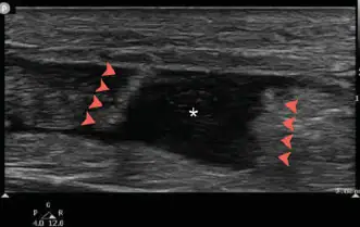
The correct diagnosis in this case is complete Achilles tendon rupture. POCUS shows tendon segment retraction (arrowheads) and fluid between the segments (asterisk). This classically occurs after a sudden forceful push-off, often during sports, with acute posterior ankle or calf pain and an audible or palpable “pop.” However, presentations may be subtle, particularly when patients have preceding tendinopathy or subacute tendon pain before complete rupture. Misdiagnosis of an ankle sprain or calf strain may occur when pain partially improves or patients retain some plantarflexion through accessory muscles.
Discussion
This case highlights an important urgent care clinical pearl: Achilles tendon rupture should remain on the differential for posterior ankle pain or ecchymosis, loss of plantarflexion, or inability to perform a single-leg heel raise, even when the Thompson test is equivocal. Recent fluoroquinolone exposure further increases clinical suspicion. Other reported risk factors include steroid use, oral bisphosphonate use, male sex, obesity, diabetes mellitus, chronic kidney disease, hyperparathyroidism, and intermittent athletic activity in deconditioned individuals.1
POCUS can rapidly confirm the diagnosis at the bedside. A high-frequency linear transducer should be used to evaluate the Achilles tendon in long- and short-axis views, scanning from the myotendinous junction to the calcaneal insertion. A normal tendon appears as a continuous fibrillar band, whereas a complete rupture demonstrates loss of fibrillar continuity, tendon retraction (arrowheads), and a fluid-filled gap (asterisk). For complete Achilles tendon rupture, ultrasound has a reported sensitivity of 94.8% and specificity of 98.7%.2
Partial tears are more challenging and may require comparison with the contralateral side or advanced imaging.3 Dynamic assessment with a real-time Achilles ultrasound Thompson test may also support the diagnosis when differentiating partial from complete rupture.4 The retrocalcaneal bursa should not be mistaken for a tendon rupture, as it may contain fluid in cases of bursitis.
Once a complete rupture is identified, or when clinical suspicion remains high despite an equivocal study, the patient should be splinted in plantarflexion, kept non-weight bearing, and referred urgently for orthopedic evaluation.
What To Look For
- In complete tears, the tendon defect is usually readily apparent, with discontinuity of the Achilles tendon fibers and retraction of the tendon ends.
- Look for anechoic or hypoechoic fluid within the defect, consistent with hemorrhage.
Pearls For Urgent Care Management
- POCUS is easy to perform and highly sensitive for complete Achilles tendon ruptures, helping to quickly make the diagnosis.
- Scan the entire tendon in both the long- and short-axis views.
- Partial tears are more subtle. Compare with the contralateral tendon and use dynamic maneuvers to improve diagnostic accuracy.
- Patients with current or recent fluoroquinolone, steroids or oral bisphosphonate use are at increased risk for Achilles tendon rupture.
References
- Xergia SA, Tsarbou C, Liveris NI, et al. Risk factors for Achilles tendon rupture: an updated systematic review. Phys Sportsmed. 2023;51(6):506-516.
- Aminlari A, Stone J, McKee R, et al. Diagnosing Achilles Tendon Rupture with Ultrasound in Patients Treated Surgically: A Systematic Review and Meta-Analysis. J Emerg Med. 2021;61(5):558-567.
- Fenech M, Ajjikuttira A, Edwards H. Ultrasound assessment of acute Achilles tendon rupture and measurement of the tendon gap. Australas J Ultrasound Med. 2024;27(2):106-119.
- Griffin MJ, Olson K, Heckmann N, Charlton TP. Realtime Achilles Ultrasound Thompson Test for the Evaluation and Diagnosis of Acute Achilles Tendon Ruptures. Foot Ankle Int. 2017;38(1):36-40.
Acknowledgement: Case provided by Tatiana Havryliuk, MD, an emergency physician based in New York, New York, and the founder of Hello Sono.
Case and images provided courtesy of Hello Sono (www.hellosono.com).
Read More
- Ruptured Baker Cyst is an Uncommon Complication of a Common Diagnosis: A Case Report
- Utility of POCUS in Skin and Soft Tissue Infection
- 51-Year-Old With Chest Pain After a Skiing Fall