Published on
Urgent message: Fractures of the distal radius are a common presentation in the urgent care setting. Nonetheless, a high index of suspicion is required for the diagnosis of a Galeazzi fracture–dislocation.
By Heather L Hinshelwood, MD and David Caro, MD
Introduction
The anatomic structure of the forearm typically maintains the integrity of the distal radioulnar joint when the radius is fractured. However, in certain situations this joint may be compromised with certain rotational forces.
Case
A 14-year-old African-American male was struck by a motor vehicle while walking. The patient was unsure as to where he was struck, how he landed, and whether or not he lost consciousness. He sustained multiple lacerations of his mid-face, left shoulder, and right lower extremity; several of these wounds required suturing. He was observed overnight in the pediatric trauma unit and discharged the next day.
Two days later, the patient returned complaining of left wrist and forearm pain. On exam, he was afebrile, and vital signs were within normal limits. The facial and extremity lacerations were healing well without evidence of infection.
The patient’s left wrist was mildly swollen and tender to palpation over the distal radius and ulna, and over the dorsal, distal radial-ulnar joint (DRUJ) region. He had no tenderness to palpation in his fingers, hand, snuffbox, elbow, humerus, or shoulder. His pulses were normal and sensation was intact. His left elbow and shoulder had full, painless range of motion. He was able to move his wrist through a full range of motion in all directions without assistance. The remainder of his exam was normal.
Plain radiographs of the patient’s left wrist and forearm revealed a non-displaced unicortical fracture of the distal third of the radius (Figures 1a and 1b). The growth plates were still open and no ulnar fracture was seen. Due to the tenderness over the DRUJ, the diagnosis of a Galeazzi fracture with some degree of disruption of the ligaments maintaining the DRUJ was entertained.
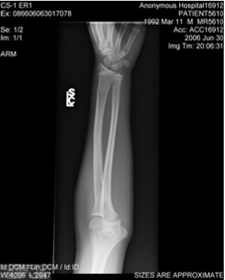
Figure 1a. Posteroanterior view of forearm. The radial fracture is unicortical and there is no shortening of the shaft.
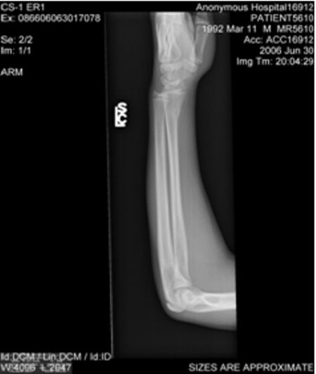
Figure 1b. Lateral view of forearm. Note that the overlap of the radius and ulna appears to be compromised.
Discussion
A Galeazzi fracture is a distal radius fracture with disruption of the radioulnar joint. The majority of the fracture sites occur between the radial insertions of the pronator teres and pronator quadratus.1
The DRUJ disruption occurs through interruption of the articular disc and the distal dorsal and volar radioulnar ligaments, especially the triangular fibrocartilage. This disruption makes for an unstable fracture, as the brachioradialis, pronator quadratus, thumb abductors and extensors, and the weight of the hand act on the distal portion of the radius and therefore distract it.2
A Galeazzi equivalent may occur in children. A distal ulnar physis fracture can occur due to the relative weakness of the growth plates. The DRUJ is not technically disrupted, but is functionally disrupted due to the loss of its anchoring on the main body of the ulna.2
Pathophysiology
Extreme pronation and extension transmit a great amount of force across the wrist and result in a fracture and shortening of the distal radius. This precedes dissociation between the distal radius and ulna, leading to the disruption of the connecting ligaments.3 Morrissy, et al noted that this pattern of injury occurs in one of two ways: either the hand is fixed and the body is rotating (e.g., an active fall on an out-stretched hand) or the body is fixed and the hand is rotating (e.g., getting a hand caught in a piece of machinery).4 Both mechanisms involve extreme extension and pronation of the wrist with rotational and fixed components.4
The radius and ulna have a mechanical dependency on one another. The lengths of the radius and ulna are roughly equal and the bones are kept in close proximity by the interosseous membrane and complex proximal and distal ligament structures. If the length of one bone is altered, there must be a disruption of the interface between the radius and ulna either proximally or distally.
Two of the more common forearm fracture–dislocations are the Monteggia and the Galeazzi. An ulnar fracture which results in proximal radioulnar dislocation is called a Monteggia fracture–dislocation. A radial fracture which results in distal radioulnar dislocation is called a Galeazzi fracture–dislocation.5
The DRUJ is maintained by the interosseous membrane connecting the radius and ulna, the pronator quadratus, and the ligaments of the wrist.3 The degree of disruption of the ligaments maintaining the DRUJ is determined by the degree to which the radius shortens.
Multiple ligaments of the wrist play an important role in maintaining this delicate architecture. The most important of these is the triangular fibrocartilage, which contains a meniscus structure that rests between the distal intersection of the radius and ulna. Disruption of the triangular fibrocartilage only may lead to a more subtle Galeazzi fracture–dislocation whose diagnosis requires a high index of suspicion. Disruption of the interosseous membrane produces the classic Galeazzi fracture–dislocation that is, typically, quite obvious.
The combination of radial shortening and ligamentous disruption makes for an unstable wrist joint.
Essentials of Clinical and Radiologic Diagnosis
The classic Galeazzi fracture–dislocation will produce an impressive clinical triad:
1. Concave deformity on the radial aspect of the wrist resulting from the fracture of the radius itself with the overall mechanical forces pulling the distal end radially.
2. Swelling and tenderness over the DRUJ due to soft tissue injury.
3. Complete radioulnar dislocation with the widening of the DRUJ and dorsal prominence of the distal ulna. The ulna may or may not have broken through the skin.1
Radiologic diagnosis
There are four basic radiologic findings that could indicate a Galeazzi fracture–dislocation (Figure 2):
1. Fracture at the base of the ulnar styloid, which is the functional equivalent of triangular fibrocartilage rupture.
2. AP widening of the DRUJ. There is no absolute width that represents a dislocation. Comparison films from the uninjured wrist might be helpful.
3. Disassociation of the radius and ulna on a lateral wrist film. The distal portions of both forearm bones no longer long lie in the same vertical plane.
Note: a true lateral film must be obtained. Any deviation from an exact, 90° true-lateral film could result in misdiagnosis (Figure 2).
4. There is ≥5 mm radial shortening (again, compare with the opposite forearm to correct for natural variation).
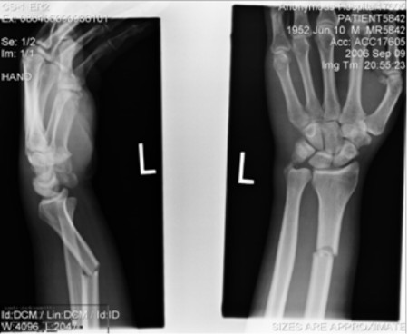
Figure 2. Lateral and AP x-rays of a Galeazzi fracture. The radial fracture in view is the first clue. The distal portions of the radius and ulna no longer overlap on the AP or lateral views. The lateral view reveals the shortening from the radial shaft fracture A high suspicion for Galeazzi fracture–dislocation injuries should be maintained in the presence of distal radial fracture, even in the absence of a protruding distal radius, concavity over the distal radius, or inability to palpate a disassociation between the distal radial and ulna. The true incidence of this injury varies from study to study. Many authors believe it to be under-diagnosed—and, therefore, under-reported.
Acutely, correct diagnosis of a more subtle Galeazzi injury may be missed by a low index of suspicion, swelling that obscures more discreet physical findings of DRUJ subluxation or dislocation, an assumption that pain is the reason for limited pronation, and attributing ulnar displacement to the position of the wrist during lateral radiography.4 Comparing both wrists is essential during the physical exam, as this may show readily apparent asymmetry from ulnar displacement.4 Also, in the presence of DRUJ disruption, wrist flexion and extension is typically not nearly as painful as attempted wrist pronation and supination, which is where the heart of the dysfunction is.4
Management Strategies
Immediate management requires a long-arm splint with the wrist in full supination, which will subsequently be converted to a cast for six to eight weeks, whether the patient receives open or closed reduction. This is necessary to approximate the DRUJ ligaments to allow appropriate healing.2,5,7 An orthopedic consult is necessary to arrange disposition.
Surgical fixation of a Galeazzi fracture–dislocation is frequently required to ensure that anatomic reduction is maintained. In Mikic’s paper, half of his adult patients (34 of 86) with the classic Galeazzi lesion were treated with closed reduction only and 80% of those had “poor” outcomes (persistent pain, forearm deformity, non-union, significant angulation or shortening of the radius, chronic dislocation of the DRUJ, restriction of pronation-supination >45°, and excessive restriction of function of the elbow and/or wrist).3
Surgical reduction is rarely required in children, however. Typically, good anatomic reduction can be achieved with closed reduction. The tough, pliant periosteum of childhood is rarely ruptured, precluding the need for indwelling hardware to maintain the reduction.3
Teaching Points
• Isolated radius fractures are more common than radial fractures with DRUJ disruption.
• This injury occurs during extremes of wrist extension and pronation.
• Presentation of Galeazzi fractures can vary from subtle physical findings that may not be readily apparent, to very obvious findings that include a pronated wrist with a limp hand and a dorsally protruding distal ulna.
• Management of this fracture in the acute setting includes closed reduction and long-arm splinting in supination. A long-arm cast will be applied for a minimum of six weeks by the orthopedic consultant.
• Non-diagnosis can lead to long-term functional impairment of the affected wrist, most notably chronic pain and limited pronation and supination.
Case Conclusion
While some portions of this patient’s exam may have been concerning for Galeazzi fracture, others were not. He did have pain on palpation of his DRUJ. However, his fracture was unicortical and non-displaced (i.e., no shortening of the radius in relationship to the ulna), he had full range of motion in all planes once his pain was controlled, and he had none of the radiographic findings indicative of DRUJ disruption (i.e., AP joint space widening, non-overlap of the radius and ulna on lateral). Conservative treatment dictated a long-arm splint in full supination.
The patient was referred to a pediatric orthopedist for definitive care.
References
1. Peimer CL, ed. Surgery of the Hand and Upper Extremity. New York: McGraw-Hill; 1996.
2. Green NE, Swiontkowski MF, eds. Skeletal Trauma in Children. 3rd ed. Philadelphia: Saunders; 2003.
3. Mikic ZD. Galeazzi fracture–dislocations. J Bone Joint Surg. 1975;57(8):1071-1080.
4. Morrissy RT, Nalebuff EA. Dislocation of the distal radioulnar joint: Anatomy and clues to prompt diagnosis. Clin Orthop Relat Res. 1979;144:154-158.
5. Reckling FW. Unstable Fracture–Dislocations of the Forearm (Monteggia and Galeazzi lesions). J Bone Joint Surg. 1982;64(6):857-863.
6. Letts M, Rowhani N. Galeazzi–equivalent injuries of the wrist in children. J Pediatr Orthop.1993;13(5):561-566.
7. Rodriguez-Merchán EC. Pediatric fractures of the forearm. Clin Orthop Relat Res. 2005;432:65-72.