Published on
Differential Diagnosis
- Acute compartment syndrome
- Cuboid fracture
- Cuneiform fracture
- Lisfranc fracture dislocation
Diagnosis
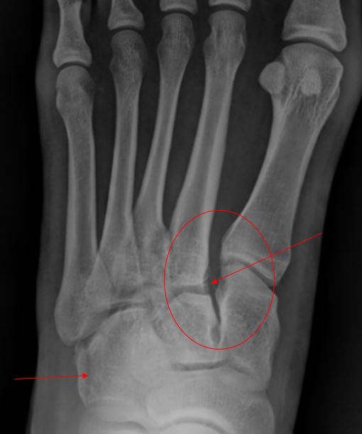
This patient was diagnosed with a Lisfranc fracture dislocation and a longitudinal cuboid fracture. Lisfranc fracture dislocation is a term that describes fractures and dislocations that occur at the junction between the tarsal bones of the midfoot and the metatarsals of the forefoot. The x-ray above shows a widening of the space between metatarsal 1 and metatarsal 2 and a widening of the space between cuneiform 1 and metatarsal 2. Named after Jacques Lisfranc, a field surgeon in the French army under Napoleon, the original context was as a new technique for amputation used to treat frostbite of the forefoot in soldiers on the Russian front.
Learnings/What to Look for
- Lisfranc fracture dislocations are most likely to occur while playing a sport, as the result of a motor vehicle accident, or during a fall from a height (such as while walking down steps or off a curb—or falling from a rock)
- Clinical findings include pain at the tarsal-metatarsal joints, swelling, ecchymosis, and potential joint instability
Pearls for Urgent Care Management
- Weightbearing x-rays should be considered to determine joint stability and presence of displacement
- Nondisplaced injuries may be treated conservatively (non-weightbearing with immobilization in a boot or short leg cast for 6 weeks, followed by progressive weightbearing)
- Displaced Lisfranc injuries are likely to require closed or open surgical reduction
Acknowledgement: Images and case presented by Experity Teleradiology (www.experityhealth.com/teleradiology).
View Similar Cases
- A 21-Year-Old Male With Foot Pain
- A Construction Worker With Sudden Foot Pain After Stepping Into A Hole