
Published on
Download the article PDF: Clinical Image Challenges September 2026
Differential Diagnosis
- Acute coronary syndrome
- Left posterior fascicular block
- Dextrocardia
- Limb lead reversal
- Marked (extreme) right axis deviation
Discussion
The diagnosis in this case is limb lead reversal. The ECG shows a regular rhythm with visible P waves before each QRS complex, consistent with sinus rhythm. There are deep S waves and negatively deflected P waves in Lead I as well as a globally positive QRS complex in aVR. These findings should raise suspicion for a limb lead reversal, specifically right arm-left arm (RA–LA) reversal, which is the most common type of electrode misplacement.1
The ECG changes seen with RA–LA reversal can be understood as a 180° horizontal rotation of Einthoven’s triangle around the aVF axis.2 This results in inversion of leads I, swapping of aVR and aVL, an apparent marked right axis deviation, and swapping of leads II and III (Figure 2).3-4 In addition, in limb lead reversal, the precordial leads typically retain normal morphology and R-wave progression, as chest lead placement remains unaffected. Electrode misplacement can lead to significant ECG misinterpretation and may result in unnecessary cardiac workup in patients without true pathology.5
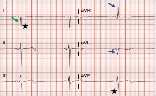
A clinician may mistake this ECG for acute coronary syndrome given the T-wave inversions in leads I and aVL and the biphasic appearing T-waves in inferior and precordial leads. However, acute coronary syndrome would not cause global inversion of lead I along with a positive aVR. This pattern should alert the clinician to repeat the ECG and confirm proper lead placement prior to making an interpretation. Additionally, there are no acute ST elevations or ST depressions in contiguous leads, ruling out ST-segment elevation myocardial infarction.
Left posterior fascicular block may be considered, as the QRS complexes in leads I and aVL are negatively deflected, leading to the appearance of a marked right axis deviation. However, left posterior fascicular block does not cause the P wave in lead I to be inverted. Additionally, isolated left posterior fascicular block is rare in the general population, making this diagnosis less likely.6
RA-LA limb lead reversal can be mistaken for dextrocardia, as both can present with an inverted lead I and a positive aVR. However, the key distinguishing feature is the R-wave progression in the precordial leads. In dextrocardia, there is reversed R-wave progression, with leads V3-V6 showing predominantly negative QRS complexes.7 In contrast, RA-LA limb lead reversal affects only the limb leads, so the precordial leads remain normal, as seen in this patient’s ECG. This finding supports limb lead reversal as the correct diagnosis.
Marked (extreme) right axis deviation may be considered given the negative QRS complexes in lead I and aVF. However, limb lead reversals are known to mimic axis abnormalities and true marked right axis deviation does not cause global inversion of lead I or a positive aVR. Therefore, this ECG does not reflect true physiological axis deviation. Clinicians should consider repeating the ECG and confirm that all leads are placed in their proper position.
What To Look For
- The key ECG clues for RA-LA lead misplacement include positive P wave, QRS complex, and T wave in aVR, apparent marked right axis deviation, and global inversion of the P wave, QRS complex, and T wave in lead I.
- If limb lead reversal is suspected, repeat the ECG and confirm proper lead placement before making an interpretation.
Pearls For Initial Management, Considerations For Transfer
- Limb lead reversal can mimic myocardial infarction and other serious pathologies. Clinical judgement is essential, and providers should have a low threshold to repeat the ECG when findings do not match the clinical picture.
- If concern for acute coronary syndrome persists after correcting lead placement, then escalating transfer to an emergency department is imperative. Recognizing limb lead reversal can help avoid unnecessary workup and inappropriate transfers.
References
- Ramadurai S, Varadarahan V, Lasrado AA, et al. A study of the Frequency of Lead Reversal at a Tertiary Care Institution. Cureus. 2025;17(9).
- Pérez-Riera AR, Barbosa-Barros R, Daminello-Raimundo R, de Abreu LC. Main artifacts in electrocardiography. Ann Noninvasive Electrocardiol. 2018 Mar;23(2)
- Limb Lead Reversal. ECG Stampede. https://www.ecgstampede.com/glossary/limb-lead-reversal/. Accessed April 8, 2026.
- Rosen AV, Koppikar S, Shaw C, Baranchuk A. Common ECG Lead Placement Errors. Part I: Limb Lead Reversals. Int J Med Students. 2014 Jul-Oct;2(3):92-8.
- Roy SK, Shah SU, Villa-Lopez E, Murillo M, Arenas N, Oshima K, Chang RK, Lauzon M, Guo X, Pillutla P. Comparison of electrocardiogram quality and clinical interpretations using prepositioned ECG electrodes and conventional individual electrodes. J Electrocardiol. 2020 Mar-Apr;59:126-133.
- Pérez-Riera AR, Barbosa-Barros R, Daminello-Raimundo R, de Abreu LC, Tonussi Mendes JE, Nikus K. Left posterior fascicular block, state-of-the-art review: A 2018 update. Indian Pacing Electrophysiol J. 2018 Nov-Dec;18(6):217-230
- Paul A, Jacob JR. Electrocardiographic lead reversals. Indian Pacing Electrophysiol J. 2023 Nov-Dec;23(6):205-213.

Read More