
Published on
Download the article PDF: Clinical Image Challenges November 2026
Differential Diagnosis
- Hypokalemia
- Hypocalcemia
- Brugada syndrome
- Left ventricular hypertrophy with strain
- Normal sinus rhythm
Diagnosis
The diagnosis in this case is hypokalemia. The ECG reveals a sinus rhythm with marked sinus arrhythmia and a ventricular rate of 64 beats per minute. There is marked prolongation of the QT interval due to the presence of prominent U waves that have fused with the T waves, creating a wide, wavy repolarization pattern across the precordial leads.
Discussion
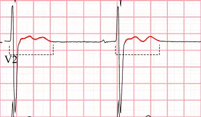
Hypokalemia is a common and potentially life-threatening electrolyte abnormality that delays ventricular repolarization.1 The characteristic ECG changes in hypokalemia include T wave flattening or inversion, ST-segment depression, and the development of prominent U waves. In severe cases such as this one, these U waves merge with the preceding T waves, creating a biphasic TU fusion complex that prolongs the QT (or QU) interval.2 This is illustrated in Figure 2.
This QU pattern in hypokalemia is caused by a delay in the repolarization phase (phase 3) of the cardiac action potential. This patient’s clinical presentation of generalized weakness combined with a QTc of >500 milliseconds and prominent precordial U waves is highly diagnostic of severe hypokalemia. Hypokalemia commonly occurs due to gastrointestinal losses (such as vomiting or diarrhea), renal losses (associated with diuretic use or hyperaldosteronism), alcohol abuse, or poor nutritional intake. It is estimated that approximately 3% of hospitalized patients have hypokalemia, and severe hypokalemia (potassium <2.5 milliequivalents per liter) is associated with a high incidence of cardiac arrhythmias.3
A long QT or QU interval increases the risk of a new electrical impulse firing before the heart has finished repolarizing – referred to as an early afterdepolarization. If premature depolarization occurs, it can trigger a life-threatening arrhythmia called torsades de pointes. Bradycardia exacerbates this risk by lengthening the repolarization phase, making overdrive pacing—either pharmacologic or electrical—a key strategy for preventing torsades de pointes in patients with acquired long QT states.
Urgent care management of suspected severe hypokalemia focuses on screening for complications, initiating replacement therapy, and immediate transfer. A thorough history should screen for symptoms of hypoperfusion, palpitations, or syncope. Initial steps include establishing intravenous access, placing the patient on continuous cardiac monitoring, and checking a basic metabolic panel to determine the potassium level. Since hypokalemia is frequently accompanied by hypomagnesemia, a magnesium level should also be checked and replaced as necessary. Patients with a severely prolonged QT or QU interval (>500 milliseconds) or who are symptomatic require immediate transfer via emergency medical services to a facility with telemetry monitoring for repletion and continuous cardiac monitoring.
Hypocalcemia prolongs the QT interval by extending the plateau phase (phase 2) of the cardiac action potential (lengthening the ST segment but with a normal T wave). In terms of the differential diagnoses, Brugada syndrome is characterized by a pseudo-right bundle branch block and downsloping ST-segment elevation in leads V1 and V2, which are absent here. Left ventricular hypertrophy with strain shows high QRS voltages and asymmetric T wave inversions in the lateral leads, and a normal sinus rhythm would not feature these severe repolarization abnormalities.
What To Look For
- Look for a wide, wavy repolarization pattern across the precordial leads, indicating the fusion of T waves and prominent U waves of severe hypokalemia.
- Bradycardia in the setting of a prolonged QT or QU interval (>500 milliseconds prolonged QT or QU interval) increases the risk of triggering torsades de pointes.
- Hypokalemia and hypomagnesemia commonly coexist, and both require aggressive replacement to stabilize ventricular repolarization.
Pearls For Initial Management, Considerations For Transfer
- Establish intravenous access and initiate continuous cardiac monitoring.
- Obtain emergency laboratory testing, including a basic metabolic panel and magnesium level if available.
- Replenish both potassium and magnesium, using oral and parenteral routes depending on patient tolerance and severity.
- Transfer the patient immediately via telemetry-monitored transport to the nearest emergency department if the QT or QU interval is >500 milliseconds or if the patient is symptomatic.
References
- Cooper B, Reynolds C. ECG Stampede: The Textbook. 3rd ed. Houston, TX: ECG Stampede Publishing; 2024.
- Cooper B, Reynolds C. ECG Stampede: The Workbook. 2nd ed. Houston, TX: ECG Stampede Publishing; 2024.
- Surawicz B, Childers R, Deal BJ, et al. AHA/ACCF/HRS Recommendations for the standardization and interpretation of the electrocardiogram: part III: intraventricular conduction disturbances. J Am Coll Cardiol. 2009;53(11):976-981. doi:10.1016/j.jacc.2008.12.013
