Published on
Download the article PDF: Clinical Image Challenges July August 2026 1
Differential Diagnosis
- Isolated malleolar fracture
- Unstable ankle fracture
- High-grade ankle sprain
- Peroneal tendinopathy
Diagnosis
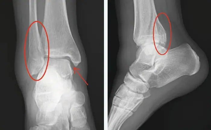
The diagnosis of an unstable ankle fracture is confirmed by radiographic findings. Images show a minimally displaced, syndesmotic spiral fibula fracture with subtle posterior malleolus involvement and possible mortise widening. This is consistent with a Weber B/C type injury. This mechanism typically involves forced supination of the foot and external rotation of the talus, often associated with ligamentous injury (e.g., anterior talofibrular ligament or deltoid ligament). Instability is present if 2 or more significant injury sites exist. Bimalleolar and trimalleolar (including the posterior malleolus, seen best on lateral x-ray views) fractures are inherently unstable and require specialist referral for orthopedic surgical management.
Ligamentous injury must also be evaluated by checking the medial clear space (talus to medial malleolus) on x-ray. A medial clear space >4 mm on standard or weight-bearing views suggests deltoid ligament disruption. Weight-bearing radiographs require ≥50 body weight and are the gold standard for assessment. Bearing weight on the ankle is often intolerably painful after the initial injury; gravity stress views are an appropriate alternative. Magnet resonance imaging/ultrasound may further delineate deltoid ligament integrity, though they do not always determine stability. Complications of unstable ankle fractures can include acute compartment syndrome as well as chronic pain and instability.
What to Look For
- Since these are often high-impact mechanisms of injury, evaluate for other injuries to the lumbar spine, head, hip or knee.
- Given the higher risk of complications from unstable ankle fractures, include a thorough neurovascular exam.
Pearls For Urgent Care Management
- Emergency conditions such as open fracture or neurovascular impairment require immediate surgical consultation and treatment in an emergency department.
- Urgent ortho consult and/or referral is recommended. Counsel patient that surgical internal fixation will likely be required if ankle is unstable.
- Acute management includes short-leg posterior splint immobilization in neutral position at 90 degrees; sugar-tong splint can be added for additional mediolateral support.
- Counsel patient to maintain non-weight-bearing status until orthopedic follow-up.