Published on
John J. Koehler MD, ABPM(OM)
I trained in Emergency Medicine in the 1980s and learned to use a slit lamp as an intern; it’s a skill that continues to serve me to this day. We had “opti-spuds” to remove foreign bodies and “opti-burrs” to debride rust rings. This was considered a core skill in my training. This is why, when I started Physicians Immediate Care in 1987, I bought a slit lamp as well as opti-spuds and opti-burrs for our first urgent care center.
Since then, our providers throughout out our many clinics have removed thousands of corneal foreign bodies and rust rings. Despite performing so many of these procedures, we have never had a single lawsuit or negative outcome. And we follow up on all these eye injuries the next day and continue see them until released.
Some physicians have opined to me that caring for corneal trauma may no longer be the standard of care for urgent care centers. In response, I have conducted a literature search and have found support for both corneal foreign body removal and rust ring debridement in the outpatient setting, including urgent cares and without obligatory ophthalmological referral.1-5
Since discomfort with these procedures seems to be increasingly common among UC providers, I wanted to discuss the relevant anatomy and procedures in the hopes of empowering us to reclaim these procedures that we can safely and effectively manage in our centers.
Basic Anatomy and Pathophysiology
The cornea is comprised of five layers, has no blood vessels, and is the most richly innervated tissue in the body.6 It also provides approximately 2/3 of the optical power of the eye.
- The epitheliumserves as the first barrier of defense. The epithelial cells constantly shed and are rapidly replaced, hence the speed with which corneal injuries will heal.7
- Bowman’s layer, the next layer, is composed of layered collagen fibers.
- Below, the stroma comprises 90% of the cornea’s thickness and provides shape and strength to the globe.
- Descemet’s membrane is an additional strong but thin sheet of tissue.
- Finally, the endothelium primarily maintains the hydrostatic balance between the cornea and the aqueous humor.
Patients with centrally located abrasions or foreign bodies (FB) often present earlier than patients with more peripheral injury, due to the increased nerve density over the visual axis. This is why such injuries often present prior to rust ring development. This greatly mitigates concern about central corneal staining and vision impairment.
Peripheral FBs (outside the visual axis), conversely, often present in a delayed fashion after a rust ring develops, as the nerve density and pain experienced is far less in this region of the cornea. Commonly, these patients will discover the FB/rust by looking in the mirror or due to minor discomfort from corneal inflammation.8 Rust can form as quickly as 4 hours after injury.9
Any nonvisual axis rust ring can be treated with confidence in our setting. The cornea usually heals in 12-48 hours from abrasions, FB removal, and/or rust ring debridement.5,9-12 Therefore, a next-day rechecks is advisable.10,13,14
Key History Features
- When did the injury occur? The longer the time since injury, the more the rust can be generated.
- What was the mechanism of injury? “Metal-on-metal” (eg, grinding-related) is specifically concerning. This mechanism can generate a high-speed fragment with the necessary velocity for globe penetration.9,15,16
- Is vision altered? Any reported loss of vision or blurriness compared to baseline is concerning for a more complicated injury.
Key Exam Features
- Visual acuity in both eyes should be obtained. This is the “vital sign of the eye.” Loss of visual acuity in the affected eye is concerning. If metal-on-metal injury occurred and their vision is reduced, x-rays (AP and lateral) of the globes should be obtained to evaluate for intra-ocular metallic FB.
- Cornea exam should be performed under magnification with either a head-mounted or slit lamp microscope. This should be done without fluorescein first, then with fluorescein to identify foreign body, rust ring, opacity, and/or leaking of intraocular contents (Seidel sign).9
- Inspect the anterior chamber for hyphema.
- Note conjunctival injection, discharge, hematoma, etc.
- Upon fluorescein staining, if you see a “rivulet” (Seidel sign), globe perforation is likely.9 Globe perforation is always an ocular emergency and requires immediate escalation of care to reduce risk for endophthalmitis and permanent visual loss. These patients should be referred to an emergency department or, if possible, for same-day ophthalmologist evaluation.
Studies
- Plain films of the orbits (AP and lateral XR) are an appropriate initial screening test if there is concern for a metallic foreign body. CT is also reasonable, but more costly and not widely available. MRI is contraindicated, as the magnet could cause migration of the FB if the metal is ferromagnetic.
Procedure
- Anesthetize the cornea with a topical anesthetic, such as proparacaine.
- Evert the lid(s) and examine for FBs adhered to the inner surface of the lids.
- Under magnification, inspect the cornea and scleral surface for FB. Inspect the anterior chamber for hyphema, which requires emergent referral. Inappropriately managed hyphema can result in acute glaucoma, among other complications.
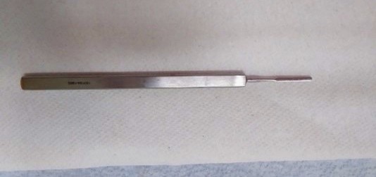
- If an FB is noted under magnification, use an Opti-Spud (Figure 1) to scoop the corneal FB off the corneal surface. If the spud is unsuccessful, you can use a splinter forceps or even the opti-burr debridement tool to loosen it from the cornea. Some metal fragments have small burrs on them that can catch onto the cornea. A bent needle (ideally 25 g) can be used if no specialized equipment is available to gently attempt dislodgement .17
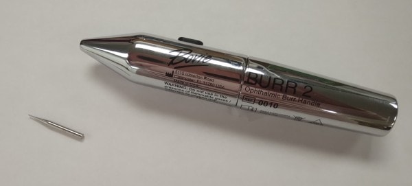
- After FB removal, if a rust ring is noted, it can be debrided with the opti-spud or opti-burr debridement tool (Figure 2. The bit spins when you compress the actuator. Fix your hand on their malar eminence and approach the rust ring tangentially and debride away the brown stain material. There is no need to place any pressure on the cornea. This procedure carries a very low risk of perforation,18,19 and is the preferred method of treatment for rust rings.9,20-23 In fact, I couldn’t find a single case in the medical literature documenting perforation has occurred using this tool.
- It is not necessary that you remove all the rust on the first visit. By the next day any residual rust will be easier to remove and closer to the surface.10
- If there is residual rust in the visual axis, then debridement should be repeated. If it is still not fully removed, refer the patient to ophthalmology to complete the debridement.
- If there is minimal residual rust outside the visual axis and the patient is asymptomatic, further debridement is not absolutely necessary. A recheck in 48 hours to confirm normal healing is reasonable.
- Tip: Practicing with these tools on a hard-boiled egg, bar of soap, or commercially available rubberized globe is a great way to gain confidence before using them on a patient’s eye.
Medications/Aftercare
- Anesthetic eyedrops are controversial, and it is not advisable to send patients home with topical anesthetics.9,10,24
- Prophylactic antibiotic eyedrops are not necessary for routine corneal abrasions and FB removal.25 They are indicated if a rust ring was present and debrided.
- Artificial tears can be dispensed to all eye injury patients.26
- Oral NSAIDs and acetaminophen are both reasonable for analgesia.
- Topical NSAIDs may slow healing and should not be prescribed.26
- Eye cover/patching is not indicated.
Work Status
- Patients often have immediate and significant benefit following FB removal and can return to regular home/work activities the same day. Contact lenses should not be applied until the cornea is healed.
Recheck
- All eye injuries should be re-evaluated the following day. Visual acuity should be rechecked at every follow-up.
- If the patient is asymptomatic on reassessment and the cornea is clear, they can be released.
- If they are asymptomatic with a small amount of residual rust outside the visual axis, one final recheck in 48-72 hours is advised.
Conclusion
It is understandable that providers often feel anxious about treating eye injuries. Many simply were not trained to do these procedures. Medical training of all varieties tends to be more focused on illness than injury. This has consequences for UC because the providers joining the work force generally have limited exposure to the procedures necessary to manage common minor injuries. Many market participants, payers, and employers have noticed that this results in a shift towards triaging patients with such minor injuries instead of simply treating them.
This is a dire problem for UC if allowed to proceed unchecked. The solution requires medical leadership to step up and teach providers the skills necessary for treating the full array of injuries for which patients might present to UC. We owe it to our communities and the employers who we serve in ensuring their work force’s occupational health is managed efficiently.
Some leaders have shared with me concerns about the time it would take their providers to treat ocular injuries. The answer, again, is not to stop doing these procedures, but again to train providers to be efficient in performing them. This comes with practice; the more your UC center sees, the better your providers will get. The literature clearly supports early removal of a corneal FB.10,13 Once trained, it only should take the provider a matter of seconds to remove a corneal FB or debride a rust ring. Referring the patient delays treatment, allows more rust to develop, and increases the risk of infection. This is the antithesis of high-quality, patient-centered care.
If we, as an industry, give up now on injury treatment (eg, lacerations, fractures, fingertip avulsions, eye injuries etc.), we will lose these patients forever. Moreover, how will we distinguish ourselves from retail and drugstore clinics? Payers are already offering $70 injury case rates because “all [we] do is refer.” Can our clinics survive if this becomes the industry standard? How many referrals of routine orthopedic cases where we could easily provide definitive care will it take for families to choose orthopedic “urgent care” centers the next time they get injured?
Furthermore, most UC websites advertise injury treatment, including fractures, yet we move closer and closer to triage instead of treatment. This is simply unfair to the patients, because they end up paying for two visits. Don’t we owe it to our communities and occ med employers that we serve to treat routine injuries?
One model to consider, if training and equipment can’t meet these needs at all your network’s centers, would be referring eye injuries, minor fractures, and fingertip injuries to one of your own clinics. This “hub-and-spoke” model allows for providers who are trained and comfortable with higher acuity to be managing these patients. It also provides better continuity.
To further demonstrate our capability in managing corneal FB and rust ring injuries, the next steps will involve studying this in our centers. Please email me at [email protected] if you’re interested in conducting a study of these injuries at your UC clinic(s).
From the beginning, our value propositionin UC has been ease of access and treatment for both minor illnesses and injuries to reduce unnecessary ED and specialist visits. As a long-time insider and stakeholder in UC, my desire is for our success and sustainability long into the future. Caring for corneal injuries is just one example of an injury we can reclaim, and with it take a big step towards securing our continued survival.
REFERENCES
- Bunuel-Jordana L, Fiore DC. Letters to the Editor: Is ophthalmologic follow-up for corneal abrasions needed? Am Fam Physician. 2004;70(1):32.
- Marx JA, Hockberger RS, Walls RM, Adams J, eds. Rosen’s Emergency Medicine: Concepts and Clinical Practice. 5th ed. St Louis, MO: Mosby; 2002:915-916.
- Tintinalli JE, Kelen GD, Stapczynski JS, eds. Emergency Medicine: A Comprehensive Study Guide. 5th ed. New York, NY: McGraw-Hill, Health Professions Division; 2000:1508-1509.
- Albert DM, Jakobiec FA, eds. Principles and Practice of Ophthalmology: Clinical Practice. Vol 5. Philadelphia, PA: WB Saunders; 1994:3384-5.
- Sabri K, Pandit JC, Thaller VT, et al. National survey of corneal abrasion treatment. Eye. 1998;12:278-281.
- Yang A Y, Chow J, Liu J. Corneal innervation and sensation: the eye and beyond. Yale J Biol Med. 2018;91(1):13-21.
- Mort RL, Douvaras P, Morley SD, et al. Stem cells and corneal epithelial maintenance: Insights from the mouse and other animal models. Results Probl Cell Differ. 2012;55:357-394.
- Ozkurt ZG, Yuksel H, Saka G, et al. Metallic corneal foreign bodies: an occupational health hazard. Arq Bras Oftalmol. 2014;77(2):81-83.
- Wilson SE, Mohan RR, Mohan RR, et al. The corneal wound healing response: cytokine-mediated interaction of the epithelium, stroma, and inflammatory cells. Prog Retin Eye Res. 2001;20(5):625-637.
- Dua HS, Forrester JV. Clinical patterns of corneal epithelial wound healing. Am J Ophthalm. 1987;104(5):481-489.
- Santen SA, Scott JL. Ophthalmologic procedures. Emerg Med Clin North Am. 1995;13(3):681-701.
- Ahmed F, House RJ, Feldman BH. Corneal abrasions and corneal foreign bodies. Prim Care Clin Office Pract. 2015;42:363-375.
- Guier CP, Stokkermans TJ. Cornea foreign body removal. StatPearls Publishing LLC. September 25, 2022. Bookshelf ID: NBK554478PMID: 32119365.
- Wilson SA, Last A. Management of corneal abrasions. Am Fam Physician, 2004;70(1):123-128.
- Pieramici DJ, Capone Jr A, Rubsamen PE, Roseman RL. Lens preservation after intraocular foreign body injuries. Ophthalmology. 1996;103(10):1563-1567.
- Woodcock MG, Scott RA, Huntbach J, Kirkby GR. Mass and shape as factors in intraocular foreign body injuries. Ophthalmology. 2006;113:2262-2269.
- Beyer H, Cherkas D. Corneal foreign body removal using a bent needle tip. Am J Emerg Med. 2012;30(3):489-490.
- Duke Elder S, ed. Textbook of Ophthalmology. Vol VI. London 1954;6125-6152.
- Foulds WS. Removal of corneal foreign bodies by disposable needle. Br Med J. 1971;3:762.
- Brown N, Clemett R, Grey R. Corneal rust removal by electric drill. Br J Ophthalmol. 1975; 59(10):586-589.
- Sigurdsson H, Hanna I, Lockwood AJ, Longstaff S. Removal of rust rings, comparing electric drill and hypodermic needle. Eye (Lond). 1987;1(Pt 3):430-432.
- Sharma S. Ophthaproblem. Can Fam Physician. 1997;43:1353-1354.
- McGuinness R, Knight-Jones D. Iron-containing corneal rust rings treated with desferrioxamine. Br J Ophthalmol. 1968;52(10):777-780.
- Shaughnessy A. Topical NSAIDs of little benefit for corneal abrasion. Am Fam Physician. 2003;67(12):2580-2584.
- Wilson SA, Last A. Management of corneal abrasions. Am Fam Physician. 2004;70(1):123-128.
- Weaver CS, Terrell KM. Evidence-based emergency medicine. Update: do ophthalmic nonsteroidal anti-inflammatory drugs reduce the pain associated with simple corneal abrasion without delaying healing? Ann Emerg Med January. 2003;41:134-40.
John J. Koehler MD, ABPM(OM) is a founding board member of the Urgent Care Association and Editor-in-Chief, OccDocOne.
