Published on
Differential Diagnosis
- ST-elevation MI (STEMI)
- Left ventricular hypertrophy (LVH) with strain
- Hyperkalemia
- Left bundle branch block (LBBB)
- Ventricular tachycardia
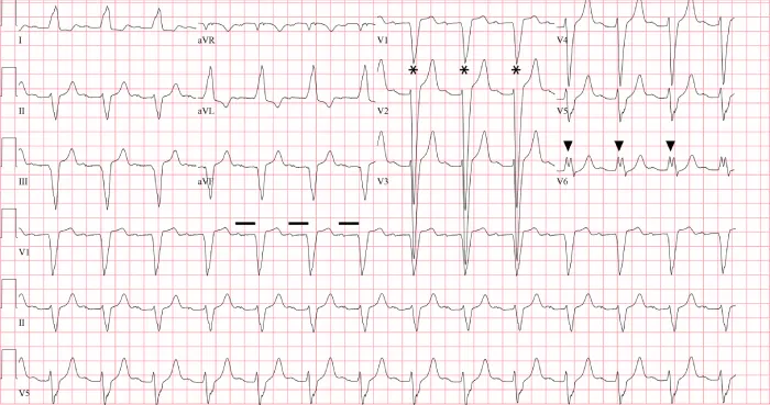
Figure 2: The wide QRS (>120 msec), dominant S wave in V1 (asterisks), broad notched R wave in V6 (arrowheads) and absent q waves in lead I, V5, and V6 indicates the presence of a left bundle branch block. The PR interval is prolonged (horizontal line).
Diagnosis
The ECG reveals a regular, wide-complex, sinus rhythm at a rate of 84 beats per minute. The wide QRS complex (>120 msec), dominant S wave in V1, broad notched R wave in the lateral leads (I, aVL, V6), and left axis deviation indicate the presence of a left bundle branch block (LBBB). The prolonged PR interval represents a first-degree atrioventricular block.
Our understanding of the trifascicular framework of the intraventricular conduction system comes from the seminal work of Rosenbaum, et al from 1969 to 1973. These works elucidated three conduction terminals—one in the right ventricle (the right bundle) and two in the left ventricle (the anterior and posterior divisions of the left bundle).1–3 Conduction disturbances of any or all three conduction terminals may result from structural abnormalities of the His-Purkinje system caused by necrosis, fibrosis, calcification, infiltrative disease, electrolyte disturbances, or impaired vascular supply.4
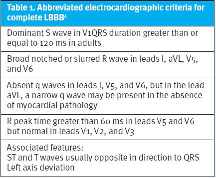
When conduction is impaired to both left ventricular terminals, the result is an LBBB. Table 1 lists the established electrocardiographic criteria for the diagnosis of LBBB.
Historically, LBBB was thought to prevent accurate recognition of acute myocardial infarction, resulting in poor allocation of reperfusion therapy.5 In fact, for many years (until 2013), new or presumed new LBBB was considered equivalent to an ST-elevation myocardial infarction.6
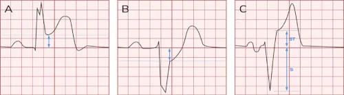
The Sgarbossa/modified Sgarbossa criteria can help to identify underlying myocardial infarction in patients with symptoms of acute coronary syndrome in the setting of a LBBB (Table 2 and Figure 3).
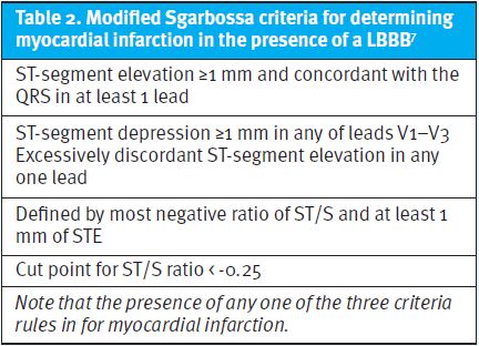
Our patient does not meet Sgarbossa criteria, but the presence of an LBBB and a first-degree atrioventricular block does indicate significant pathologic conduction disease. The symptomatic patient with an LBBB should be transferred to a catheterization-capable facility for further work-up. The ECG Stampede glossary at www.ecgstampede.com/glossary includes additional examples.
Learnings/What to Look for
- Electrocardiographic findings of left bundle branch blocks include a wide QRS, a dominant S wave in V1, and a notched or slurred R wave in leads I, aVL, V5, and V6
- Apply the modified Sgarbossa criteria for consideration of myocardial infarction in patients with symptoms of acute coronary syndrome with a left bundle branch block
- Always compare with prior ECGs when available
Pearls for Initial Management and Considerations for Transfer
- Patients with symptoms concerning for acute coronary syndrome should be transferred to catheterization-capable facility for evaluation
- A new left bundle branch block, in and of itself, does not indicate the need for emergent reperfusion; however, the provider must always consider the entire clinical picture
References
- Rosenbaum MB. The hemiblocks: diagnostic criteria and clinical significance. Mod Concepts Cardiovasc Dis. 1970;39(12):141-146.
- Rosenbaum MB, Elizari MV, Lazzari JO, et al. Intraventricular trifascicular blocks. Review of the literature and classification. Am Heart J. 1969;78(4):450-459.
- Elizari MV, Acunzo RS, Ferreiro M. Hemiblocks revisited. Circulation. 2007;115(9):1154-1163.
- Surawicz B, Childers R, Deal BJ, Gettes LS. AHA/ACCF/HRS Recommendations for the Standardization and Interpretation of the Electrocardiogram. Part III: Intraventricular Conduction Disturbances a Scientific Statement from the American Heart Association Electrocardiography and Arrhythmias Committee, Council on Clinical Cardiology; the American College of Cardiology Foundation; and the Heart Rhythm Society. J Am Coll Cardiol. Published online 2009.
- Cai Q, Mehta N, Sgarbossa EB, et al. The left bundle-branch block puzzle in the 2013 ST-elevation myocardial infarction guideline: from falsely declaring emergency to denying reperfusion in a high-risk population. Are the Sgarbossa criteria ready for prime time? Am Heart J. 2013;166(3):409-413.
- Antman EM, Anbe DT, Armstrong PW, et al. ACC/AHA guidelines for the management of patients with ST-elevation myocardial infarction – Executive summary: A report of the American College of Cardiology/American Heart Association Task Force on Practice Guidelines (writing committee to revise the 1999 guidelines for the management of patients with acute myocardial infarction). Circulation. 2004;110(5):588-636.
- Meyers HP, Limkakeng AT, Jaffa EJ, et al. Validation of the modified Sgarbossa criteria for acute coronary occlusion in the setting of left bundle branch block: a retrospective case-control study. Am Heart J. 2015;170(6):1255-1264.
Download the article PDF: A 69-Year-Old Male with Left-Sided Chest Pain and Dyspnea for 3 Days