Published on
Differential Diagnosis
- Bronchiolitis
- Pneumonia
- Stridor
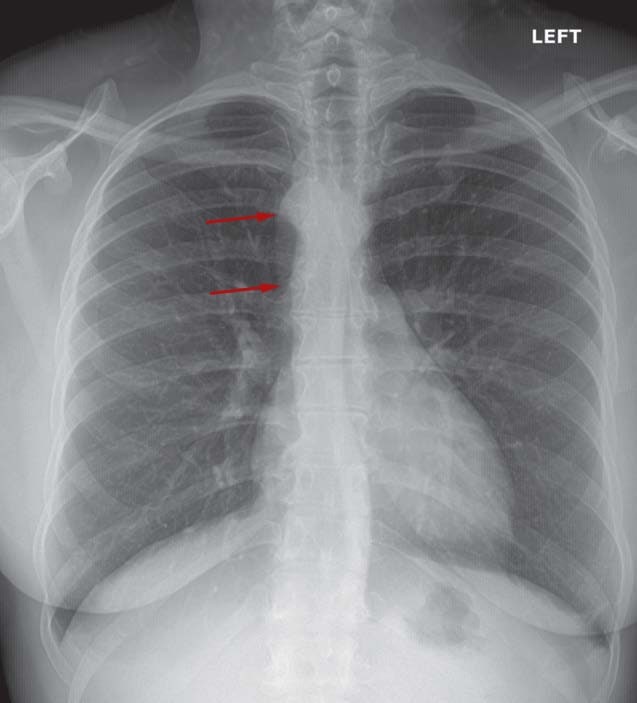
- Right aortic arch
Diagnosis
This patient was diagnosed with right aortic arch. The two most common patterns of right aortic arch are the right-sided aortic arch with mirror image branching and the right-sided aortic arch with aberrant left subclavian artery. This occurs in approximately 0.1% of the population.
Learnings/What to Look for
- Right arch with mirror image branching is associated with cyanotic congenital heart disease, including tetralogy of Fallot, truncus arteriosus, tricuspid atresia, and transposition of the great vessels
- Right arch with aberrant subclavian artery rarely produces symptoms as it usually has normal intracardiac anatomy. It is usually incidental although, rarely, it can cause esophageal and/or tracheal compression
Pearls for Urgent Care Management
- Generally, an isolated right aortic arch is a benign lesion
- Right aortic arch and left pulmonary artery anomalies may be more concerning, as well as being more difficult to identify
- Referral to cardiology is appropriate
Acknowledgment: X-ray and case presented by Experity Teleradiology (www.experityhealth.com/teleradiology).
A 35-Year-Old with a Persistent, Frequent Cough
1 2