Published on
Download the article PDF: Clinical Image Challenges July August 2026 1
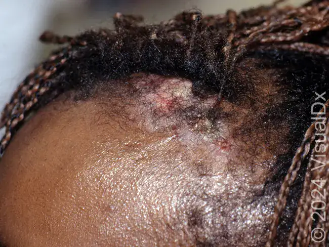
A 63-year-old woman visits urgent care with the chief complaint of an asymptomatic scabby growth on her scalp that developed 10 months prior and is gradually getting larger. The patient has no systemic symptoms and is otherwise well. She has a history of working as a welder in a metal fabrication plant for the past 35 years. On dermatological examination, an eroded and crusted plaque is seen on the frontal scalp. A punch biopsy reveals strands and cords of epithelial cells with prominent nuclear atypia, squamous pearls, and pleomorphic and hyperchromatic squamous cells with variable nuclear size.
View the image taken and consider what your diagnosis and next steps would be. Resolution of the case is described on the following page.