
Published on
Download the article PDF: Clinical Image Challenges November 2026
Differential Diagnosis
- Secondary chondrosarcoma
- Myositis ossificans
- Osteochondroma
- Parosteal osteosarcoma
- Baker cyst
Diagnosis
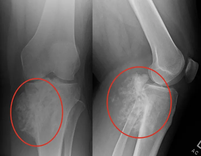
The leading diagnosis is osteochondroma. This radiograph shows a large, lobulated, sessile surface lesion arising from the proximal tibia with extensive mineralized matrix, which is consistent with a cartilage-containing surface bone tumor. There is cortical and medullary continuity between the lesion and the parent bone seen on the anteroposterior (AP) view which is a hallmark of osteochondroma. The lateral view demonstrates significant posterior extension, raising concern for possible complications, including bursa formation, fracture of the stalk, or malignant transformation.
Osteochondroma is the most common benign bone tumor, making up 20–50% of all benign bone tumors. Approximately 1% of solitary osteochondromas progress through malignant transformation to secondary peripheral chondrosarcoma. In this particular case, the large size of the mass, the history of chronic pain with acute exacerbation, and the context of the patient’s age and skeletal maturity mandate further evaluation.
What to Look For
- Red flags for potential malignancy include new-onset or worsening pain after skeletal maturity, continued growth after growth plate closure, and a cartilage cap thickness of >1.5–2 cm on magnetic resonance imaging (MRI) or computed tomography (CT), if advanced imaging is available.
- Acute exacerbation of pain may represent a fracture through the osteochondroma stalk (particularly in athletes), bursa formation over the lesion, or neurovascular compression – all recognized complications that can cause sudden worsening of symptoms without malignant transformation.
Pearls for Urgent Care Management
- Concern for malignancy indicates a need for advanced imaging. MRI with contrast is the imaging study indicated to differentiate further.
- An urgent referral to orthopedics is warranted for further workup given the presentation.
- Counsel the patient that a biopsy will likely be performed by the orthopedic specialist.