
Published on
Download the article PDF: Urgent Care Evaluation And Management Of Acromioclavicular Separation
Urgent Message: Urgent care management of acromioclavicular joint separation consists of patient education on immobilization and pain management with consideration for emergency department referral when red flags are present.
Jerome Walsh, BS; Clinton Hartz, MD; Michael B. Weinstock, MD
Keywords: acromioclavicular joint separation; ac joint injury; shoulder trauma; rockwood classification; shoulder injury; urgent care
Questions for the Clinician at the Bedside
- Which demographics are most likely to present with an acromioclavicular joint separation?
- What is the name of the classification system for acromioclavicular joint injuries?
- Which patients presenting with acromioclavicular joint separation should be referred to the emergency department?
- When would a patient with a suspected acromioclavicular joint separation require surgery?
Editor’s Note: While the images presented here are authentic, the patient case scenarios are hypothetical.
Abstract
Acromioclavicular (AC) joint separation is a shoulder injury that involves the articulation between the acromion process of the scapula and the distal end of the clavicle. Separation occurs most frequently from direct trauma, such as a fall, often experienced while playing sports or cycling. As a result of this trauma, the acromioclavicular (AC) ligament and coracoclavicular (CC) ligament may become torn, resulting in joint instability.
Diagnosis is guided by a thorough history, physical examination, and plain radiograph films. The history likely identifies the mechanism of injury as well as other potential problems. Physical examination includes inspection and palpation of the AC joint to assess range of motion and strength and to identify potential neurovascular compromise. Plain films evaluate joint alignment and classify the injury according to the Rockwood classification system, which categorizes AC separations on a scale ranging from I-VI to determine the optimal treatment. Types I-II are treated conservatively, while IV-VI are treated surgically. Type III injuries are subject to provider and patient preference.
Urgent care management of AC separation should consist of patient education on recovery, immobilization, and pain management with acetaminophen and nonsteroidal anti-inflammatory medications. The provider must also rule out red flags, and if present, refer to the emergency department.
Clinical Scenario
A 25-year-old male presented to urgent care for right-sided shoulder pain following a fall during an intramural soccer game earlier the same day. He reported sharp pain at the distal end of the clavicle that worsened with movement. He denied fever, vomiting, chest pain, shortness of breath, abdominal pain, and head trauma. On exam, the right shoulder was tender with palpation over the acromioclavicular (AC) joint, and he had full range of motion (ROM), despite feeling pain with articulatory motion. His lungs were clear to auscultation bilaterally. His heart had a regular rate and rhythm with no murmurs, gallops, or rubs. His neck had full range of motion with no pain on palpation and no swelling or discoloration.
Background and Epidemiology
Shoulder pain affects 5%–47% of adults.1 In an epidemiologic analysis of emergency departments and athletes, shoulder injuries represented 4.35% of reported injuries.2 Approximately 9% of shoulder injuries involve the AC joint.3 The majority of AC injuries occur in individuals in their 20s and are 5 times more likely to happen to men than women. Nearly all of these injuries occur during contact sports such as hockey, soccer, American football, and high velocity/impact sports such as skiing/snowboarding or mountain biking.4,5 A cohort study out of Oslo, Norway, found that among a patient population, 24% of AC injuries were due to soccer, 16% to cycling, 11% to martial arts, and 18% to skiing/snowboarding.6
Understanding the Anatomy
The shoulderjoint is composed of 3 bones: the humerus; the scapula; and the clavicle. The insertion of the humerus into the glenoid cavity of the scapula is termed the glenohumeral joint (GH). The insertion of the clavicle onto a bony process of the scapula, termed the acromion, is known as the AC joint.
The acromion and clavicle are held together by the acromioclavicular and coracoclavicular ligaments. These provide horizontal and vertical stability, respectively. During shoulder injuries, adjacent structures may be damaged including:
- The axillary nerve, which supplies the deltoid and teres minor muscles
- The axillary artery, which helps supply the entire upper extremity
- The humeral head, which sits in the glenoid cavity and is stabilized by the muscles of the rotator cuff; the rotator cuff muscles are commonly termed “SITS,” and include the supraspinatus, infraspinatus, teres minor, and subscapularis muscles7
History
An AC separation may occur from direct trauma, such as a mechanical fall, or a syncopal episode. The mechanism classically is a direct blow to the superior aspect of the joint with the arm adducted (at the patient’s side) resulting in damage to the joint and surrounding ligaments.1,4
Obtaining a detailed history of the injury from the patient is vital in determining the management of the condition. It is important to determine the onset, mechanism, location of pain, and exacerbating and relieving factors. Assessment of neurovascular compromise includes inquiring about numbness and paresthesia.4
In the absence of a clear traumatic mechanism, a clinician should consider referred pain from cardiac or abdominal/diaphragmatic etiology and inquire about chest pain/discomfort, dyspnea, diaphoresis, fever, abdominal pain, or mental status changes.8 In patients with risk factors for malignancy, targeted questions regarding history of cancer, unexplained weight loss, or other constitutional symptoms may be considered.8 Additionally, a clinician should be encouraged to rule out arthritic and rheumatological causes of pain.8
Physical Examination
Examination may include inspection for visible deformities, erythema, swelling, ecchymosis, skin dimpling, and asymmetry of the shoulder girdle when compared to the contralateral side. Skin tenting may be present over the AC joint. Palpation typically reveals point tenderness at the AC joint and may demonstrate a palpable depression between acromion and humeral head indicating hyperlaxity of the glenohumeral joint, also known as an empty sulcus sign. Active and passive range of motion should be assessed, with attention to pain during shoulder abduction, adduction, flexion, extension, internal, and external rotation.
Examination may include the O’Brien test.9 The O’Brien test is performed by flexing the shoulder to 90°, adducting it 10°–15°, and internally rotating the arm so the thumb points downward while the examiner applies a downward force to the forearm. The maneuver is then repeated with the arm in external rotation (thumb upward). If the patient feels pain with both external and internal rotation, it can indicate AC joint pathology. 9 A neurovascular examination should be performed including upper extremity distal pulses and sensation.1,4 The resting position of the arm should be noted as patients with acromioclavicular joint separation often demonstrate protective posturing of the affected extremity.4 Consideration of shoulder dislocation is also warranted; anterior dislocations present in abduction and external rotation, posterior dislocations in adduction and internal rotation, and inferior dislocations in abduction with the elbow flexed and hand behind the head.1,10
Diagnostic Testing
Radiography
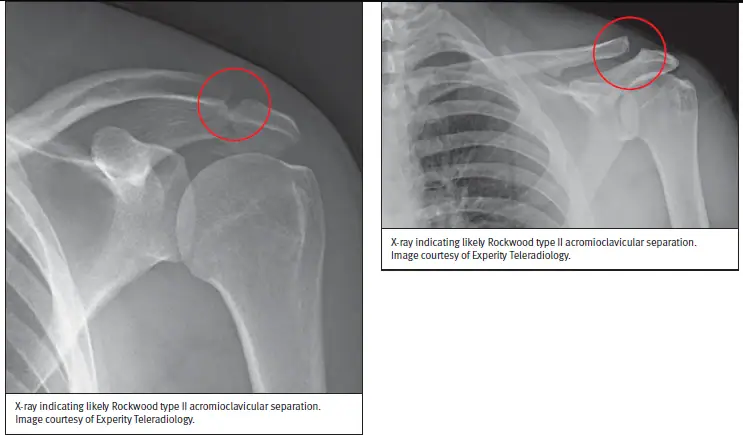
The primary imaging modality for AC separation is a plain film x-ray including anteroposterior, Zanca, and axillary views of the affected shoulder. 1,4 Findings may include widening of the joint space (Figures 1-2) or displacement of the clavicle relative to the acromion. These findings are typically compared with the contralateral shoulder to more accurately assess the extent of the injury. 1,4
The severity of the injury is commonly classified with the Rockwood classification system (Table 1).5
| Type | AC Ligament | CC Ligament | AC Joint | Deltoid and Trapezius Muscles |
| I | Sprained | Intact | Normal alignment with tenderness over AC joint | Intact |
| II | Complete Tear | Sprained; interval widened <25% | Disrupted joint space widened in transverse plane | Partial detachment possible |
| III | Complete Tear | Complete tear; interval widened 25%–100% | Dislocated with clavicle displaced superiorly | Moderate likelihood of detachment from distal clavicle |
| IV | Complete Tear | Complete tear | Dislocated with clavicle displaced posteriorly | Severe likelihood of detachment from distal clavicle |
| V | Complete Tear | Complete tear; interval widened 100%–300% | Dislocated with clavicle displaced superiorly | Severe likelihood of detachment from distal clavicle |
| VI | Complete Tear | Intact with decreased or reversed interval | Dislocated with clavicle displaced inferiorly usually in the sub coracoid position | Intact, partial, or complete detachment from distal clavicle |
Rockwood Criteria and Its Limitations
Management of acromioclavicular joint injuries is guided by the Rockwood classification. Type I and II injuries are typically treated conservatively,1,12 whereas management of type III injuries remains controversial and may involve either operative or nonoperative approaches. One study found that approximately 80% of patients with a type III injury have favorable outcomes with nonoperative treatment.1,13 However, in a follow-up study, 18–20 years postinjury, patients with Rockwood type III injuries managed nonoperatively were found to have higher rates of bony prominence and joint instability than those treated surgically.14 Surgical treatment may be appropriate in patients who require expedited return to high-demand activities such as athletes, military personnel, or manual laborers1. Type IV–VI injuries generally require surgical intervention.15
The Rockwood classification system remains the most widely used method for classifying AC joint injuries. However, alternative methods have been proposed such as the ISAKOS modified Rockwood classification and the circles measurement technique. The ISAKOS classification is used solely to further classify Rockwood type III injuries, breaking them down into 2 subtypes—type IIIA (stable) and IIIB (unstable)—to help address the controversy around management.16 However, differentiating between the 2 subtypes does require additional imaging and follow-up limiting its clinical use in an urgent care setting. The circles measurement technique compares the center-to-center distance between 2 circles drawn at the lateral clavicle and anteromedial acromion on radiographic films, then measures this distance against the same measurement on the contralateral uninjured shoulder to generate a single value. This technique has been used to classify Rockwood types II-V and has demonstrated good reliability (intraclass correlation coefficient of 0.976). Given this reliability, the circles measurement may provide additional insight into managing Rockwood injuries and may help identify patients with a type III injury who would be a candidate for surgical intervention vs conservative treatment. Despite this technique’s potential, it is not a mainstay in clinical practice.17
Advanced Modalities
If plain radiographs are inconclusive or there is concern for additional injury beyond the AC joint itself, clinicians may consider additional imaging from computed tomography (CT), ultrasound (US), or magnetic resonance imaging (MRI). US and MRI are effective at evaluating rotator cuff pathology including partial- or full-thickness tears of the rotator cuff tendons. In partial rotator cuff tears, MRI was found to be more sensitive than an ultrasound probe (74% vs 52%).18 The American College of Radiology recommends the use of CT to identify scapular fractures that may have been missed on plain radiographs.19
Urgent Care Management
Management of AC joint separation in an urgent care setting is primarily focused on determining whether the injury can be managed conservatively or requires surgical intervention. Plain film radiographic imaging plays a large role in determining the severity of the injury with treatment recommendations stemming from the Rockwood criteria.1,4
Conservative Treatment
Conservative treatment can be both nonpharmacological and pharmacological depending on patient preference. Nonpharmacological methods include ice, activity modification, and temporary immobilization with a sling and swathe for comfort.1,12 As symptoms improve, patients should begin gradual mobilization with progressive range-of-motion exercises to maintain function and facilitate recovery. Pharmacological management focuses on pain control and includes nonsteroidal anti-inflammatory medication and/or acetaminophen.10
Outpatient Referral
Most low-grade AC joint injuries have favorable outcomes with conservative management.1 Patients who remain symptomatic after 7 days of nonoperative treatment may warrant further evaluation.20 Injuries type III and higher warrant referral as they may require surgical intervention.
Indications for Referral to ED
If a patient presents with any of the following symptoms, consider referring them immediately to the nearest emergency department (ED):
- Severe pain that cannot be controlled by analgesics
- Hemodynamic instability
- Concern for open fracture, infection, or pneumothorax
- Neurovascular compromise
- Polytrauma
Red Flags and Legal Pitfalls
- Document neurovascular status.
- Obtain multiple views of the shoulder to evaluate for shoulder dislocation.
- Include acute coronary syndrome, pulmonary embolism, pneumothorax, and aortic dissection on the differential if the patient’s history, symptoms, risk factors, or mechanism of injury do not clearly support a musculoskeletal cause.
- Consider a posterior shoulder dislocation in patients whose mechanism of injury included falling onto a flexed and adducted arm, patients with seizure activity, and patients with a history of electrical shock or burn.
Clinical Scenario Conclusion
Plain radiographs at urgent care demonstrated AC joint separation consistent with a low-grade Rockwood type II injury. Given the absence of neurovascular compromise, polytrauma, or associated fracture, the injury was managed conservatively. The patient was placed in a sling and swathe and told to advance movement as tolerated. Acetaminophen and ibuprofen were recommended for pain control.
Takeaway Points
- Low-grade AC injuries diagnosed as Rockwood type I and II are managed conservatively. High-grade AC injuries diagnosed as Rockwood type IV-VI are treated surgically.
- Rockwood type III injuries are initially treated conservatively but may be considered for surgical treatment on a case-by-case basis.
- If symptoms persist for longer than 7 days, the patient may be a candidate for further evaluation.
Manuscript submitted March 27, 2026; accepted September 3, 2026.
References
- Simon LM, Nguyen V, Ezinwa NM. Acute shoulder injuries in adults. Am Fam Physician. 2023;107(5):503-512.
- Reiad TA, Peveri E, Dinh PV, Owens BD. Epidemiology of shoulder injuries presenting to US emergency departments. Orthopedics. 2025;48(2):e81-e87. doi:10.3928/01477447-20250204-03
- Chillemi C, Franceschini V, Dei Giudici L, et al. Epidemiology of isolated acromioclavicular joint dislocation. Emerg Med Int. 2013;2013:171609. doi:10.1155/2013/171609
- Albishi W, AlShayhan F, Alfridy A, Alaseem A, Elmaraghy A. Acromioclavicular joint separation: controversies and treatment algorithm. Orthop Rev (Pavia). 2024;16:94037. doi:10.52965/001c.94037
- Gorbaty JD, Hsu JE, Gee AO. Classifications in brief: Rockwood classification of acromioclavicular joint separations. Clin Orthop Relat Res. 2017;475(1):283-287. doi:10.1007/s11999-016-5079-6
- Skjaker SA, Enger M, Engebretsen L, Brox JI, Bøe B. Young men in sports are at highest risk of acromioclavicular joint injuries: a prospective cohort study. Knee Surg Sports Traumatol Arthrosc. 2021;29(7):2039-2045. doi:10.1007/s00167-020-05958-x
- Hermans J, Luime JJ, Meuffels DE, Reijman M, Simel DL, Bierma-Zeinstra SMA. Does this patient with shoulder pain have rotator cuff disease? The Rational Clinical Examination systematic review. JAMA. 2013;310(8):837-847. doi:10.1001/jama.2013.276187
- Cheung JM, Ikeda DJ, Lee CD. A 47-year-old woman with left shoulder pain. NEJM Evid. 2025;4(6):EVIDmr2500031. doi:10.1056/EVIDmr2500031
- O’Brien SJ, Pagnani MJ, Fealy S, McGlynn SR, Wilson JB. The active compression test: a new and effective test for diagnosing labral tears and acromioclavicular joint abnormality. Am J Sports Med. 1998;26(5):610-613.
- Herring SA, Kibler WB, Putukian M, et al. Initial assessment and management of select musculoskeletal injuries: a team physician consensus statement. Med Sci Sports Exerc. 2024;56(3):385-401. doi:10.1249/MSS.0000000000003324
- Williams GR Jr, Nguyen VD, Rockwood CA Jr. Classification and radiographic analysis of acromioclavicular dislocations. Appl Radiol. 1989;18:29-34.
- Ma R, Smith PA, Smith MJ, Sherman SL, Flood D, Li X. Managing and recognizing complications after treatment of acromioclavicular joint repair or reconstruction. Curr Rev Musculoskelet Med. 2015;8(1):75-82. doi:10.1007/s12178-014-9255-6
- Tamaoki MJS, Lenza M, Matsunaga FT, Belloti JC, Matsumoto MH, Faloppa F. Surgical versus conservative interventions for treating acromioclavicular dislocation of the shoulder in adults. Cochrane Database Syst Rev. 2019;10(10):CD007429. doi:10.1002/14651858.CD007429.pub3
- Joukainen A, Kröger H, Niemitukia L, Mäkelä EA, Väätäinen U. Results of operative and nonoperative treatment of Rockwood types III and V acromioclavicular joint dislocation: a prospective, randomized trial with an 18- to 20-year follow-up. Orthop J Sports Med. 2014;2(12):2325967114560130. doi:10.1177/2325967114560130
- Berthold DP, Muench LN, Dyrna F, et al. Current concepts in acromioclavicular joint instability: a proposed treatment algorithm for acute and chronic AC-joint surgery. BMC Musculoskelet Disord. 2022;23(1):1078. doi:10.1186/s12891-022-05935-0
- Rosso C, Martetschläger F, Saccomanno MF, et al. High degree of consensus achieved regarding diagnosis and treatment of acromioclavicular joint instability among ESA-ESSKA members. Knee Surg Sports Traumatol Arthrosc. 2021;29(7):2325-2332. doi:10.1007/s00167-020-06286-w
- Murphy RJ, Moor BK, Lesniewski PJ, Hayoz A, Alcantara W, Zumstein MA. Evaluation of the circles measurement and the ABC classification of acromioclavicular joint injuries. Am J Sports Med. 2021;49(6):1619-1625. doi:10.1177/03635465211003300
- Lenza M, Buchbinder R, Takwoingi Y, et al. Magnetic resonance imaging, magnetic resonance arthrography and ultrasonography for assessing rotator cuff tears in people with shoulder pain for whom surgery is being considered. Cochrane Database Syst Rev. 2013;(9):CD009020. doi:10.1002/14651858.CD009020.pub2
- Laur O, Ha AS, Bartolotta RJ, et al. ACR Appropriateness Criteria acute shoulder pain: 2024 update. J Am Coll Radiol. 2025;22(5 suppl):S36-S47. doi:10.1016/j.jacr.2025.02.015
- Saade F, Carminati F, Bouteille C, et al. Acromioclavicular joint separation: retrospective study of non-operative and surgical treatment in 38 patients with grade III or higher injuries and a minimum follow-up of 1 year. Orthop Traumatol Surg Res. 2023;109(4):103405. doi:10.1016/j.otsr.2022.103405
