Published on
Urgent message: Significant cervical spine injuries are rare in urgent care but missing one can have serious implications for patient and provider.
ERICA MARSHBURN, BS, BA, and JOHN SHUFELDT, MD, JD, MBA, FACEP
In this continuing series on back pain diagnostics in urgent care medicine, we urge providers to carefully consider any high-risk spinal conditions that could be presenting as simple back pain. Although many cases of back pain can be attributed to musculoligamentous injury and respond well to physical measures and pain medication, it is important to pay attention to signs and symptoms of more precarious spinal conditions. In order to avoid diagnostic mishaps, providers should be aware of complaints indicative of spinal injury or degeneration.
Case Presentation
A 17-year-old male presents with para-spinous cervical pain after a front-end automobile collision 3 days ago. He says he was driving and was wearing a seatbelt, and his car suffered moderate front-end damage. The patient was ambulatory on the scene and had a contusion to his forehead, but did not lose consciousness and had no chest wall pain.
Pertinent Physical Exam
All vital signs are normal. HEENT examination demonstrates ecchymosis to forehead.
The patient’s neck elicits para-spinous muscle spasm and slight midline bony tenderness. Neurological exam reveals that the central nervous system is intact with no focal neurological deficits. Chest exam indicates tenderness to the anterior chest wall, with no bruising or crepitus.
Labs/Imaging
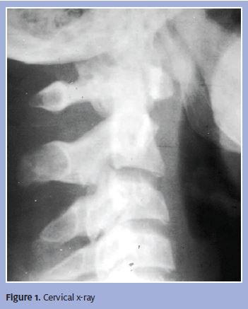
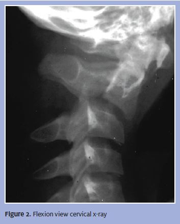
No lab tests are indicated. A plain film is ordered, revealing the images in Figures 1 and 2.
The first view shows an adolescent cervical spine, as determined by the non-fused bony end plates. These are evident at the inferior surfaces of each vertebral body. This patient’s neck was “cleared” after the first film and the patient was then allowed unprotected movement. Shortly thereafter, he developed symptoms consistent with cervical injury. If you look closely, you can see a widened pre-vertebral soft tissue space, a winded predental space, and a subtle widening of the C2/C3 interspinous space with subtle anterior subluxation of C2 on C3 or reversal of the lower cervical lordosis.
With a passive patient flexion view, we now see obvious ligaments disruption of C2-C3 (Figure 2). Widening of the interspinous space and a subluxation/dislocation of C2 on C3 are apparent.
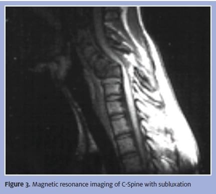
Magnetic resonance imaging (MRI) demonstrates evidence of cervical ligamentous injury and cord compression
(Figure 3).
Diagnosis
The patient was diagnosed with C2-C3 anterior subluxation. A subluxation is an aggregation of functional, structural, and pathological changes in the spinal joints that can compromise neural integrity and many affect organ system function. A lateral radiograph with the neck in neutral position may only show widening of both the interspinous and intervertebral spaces posteriorly at the level of injury. Oblique views may demonstrate widening or abnormal alignments of the facets.
Findings are often subtle and may be missed if flexion views are not obtained.
Differential diagnoses that should be considered are cauda equine syndrome, cervical strain, vertebral artery dissection, hanging injuries and strangulation, neck trauma, spinal cord neoplasm, septic shock, spinal cord infection, spinal cord injury, thoracic outlet syndrome, and torticollis.
Trauma from an automobile accident, falling, poor lifting techniques and bad posture can cause or exacerbate subluxations. Cervical subluxation is a potentially unstable condition that can cause cord compression. Cervical subluxation can cause symptoms ranging from migraines to insomnia, tingling, facial pain, dizziness, and difficulties with balance.
Course and Treatment
The patient should be referred to an emergency department for consultation with a neurosurgeon, and can be given a C-spine collar to immobilize the cervical spine. Treatment ranges from conservative rehabilitation to surgical stabilization.
Discussion
Vertebrae that are not properly aligned create tension or stretching in the nerves of the spinal cord and around the spine. Nerve signals cannot be transmitted properly in this situation, causing a number of deleterious side effects. It is important to correctly diagnose subluxations because undiagnosed spinal trauma can significantly impair sensory, motor and involuntary functions.
Imaging is very important in correctly diagnosing subluxation. Plain films have the benefit of being relatively easy to obtain, depending on patient body morphology. Also, the control of the patient and direct observation are not relinquished because most of these studies can be done in an urgent care setting.
Bony injuries and the soft tissue abnormalities associated with fractures are common with cervical subluxation. The negative predictive value of plain films, however, may not be high enough with a severe mechanism of injury. An occult fracture or ligament injury can still exist with “normal films.” Alignment or the lack thereof may be seen if ligament injuries are present. Limitations that can decrease the usefulness of plain films include patient body habitus, lack of patient cooperation, and associated musculoskeletal injuries. Further, a certain percentage of patients may have fractures or ligamentous injuries that are unidentifiable even on good quality plain films.
Plain films with supplemental stress views are very helpful to identify ligament injuries when neutral views are thought to be normal. These should only be obtained in patients who are cooperative and have no signs or symptoms of a cord injury. Obviously, that would not include neck pain because pain itself would not disqualify a patient from getting flexion or extension views.
In theory, stress views can cause cord damage, but the risk is small if these views are obtained properly. Ask the patient to flex his or her neck forward until just at the point of discomfort. Typically, that is enough forward motion to identify a ligamentous injury. Occasionally, occult fractures that are invisible on plain films may become apparent on stress views, although that is not intended purpose of the studies.
Computed tomography (CT) scans (obviously contrast is not needed for the trauma C spine) should identify all cervical fractures that are present, particularly those requiring coronal and sagittal reconstruction. Rapid-sequence scanners allow additional studies to be done with minimal time delay. If you believe a patient requires a CT scan of the C-spine to rule out an injury, sending him or her to the nearest emergency department is typically the path of least resistance.
MRI is an important outpatient imaging technique that provides objective visualization of bony and soft tissue injuries. Rarely is MRI necessary during the acute phase of an injury.
Significant cervical spine injuries are rare in an urgent care setting. That having been said, providers must remain vigilant because missed spinous injuries are a significant patient safety and medical malpractice concern.

John Shufeldt, MD, JD, MBA, FACEP