Published on
 Differential Diagnoses
- Osteolytic lesion
- Finger dislocation
- Spiral fracture of the distal phalanx
- Comminuted fracture
Physical Examination
On physical examination, the patient is alert and oriented, is not in acute distress, and is breathing comfortably. He has a temperature of 98.4°F (36.8°C), a pulse of 92 beats/min, a respiration rate of 16 breaths/min, a blood pressure of 112/76 mm Hg, and an oxygen saturation of 99% on room air.
His right middle finger has slight swelling over the DIP joint, but there are no skin color changes. When the hand is held parallel to the ground, there is a palmar droop to the distal phalanx.
The neurovascular status is intact, with good capillary refill. He experiences pain with palpation over the DIP joint, mostly dorsally. Strength testing does not result in active dorsiflexion, but the patient can actively perform palmar flexion at the DIP joint. He has no pain with palpation of the PIP joint.
Diagnosis
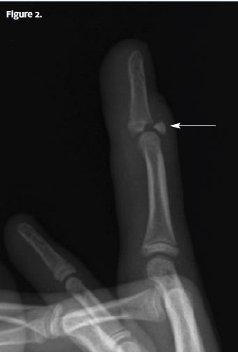
An x-ray is obtained that shows a mallet finger (Figure 2) with avulsion of the proximal aspect of the distal phalanx.
Treatment
Conservative initial treatment is the standard of care for type 1 injuries (closed injuries either with or without avulsion fracture) when there is an avulsion fracture involving less than one-third of the articular surface and no DIP joint subluxation. A splint can be applied to the DIP joint only; the PIP joint does not need to be immobilized. The DIP joint should be immobilized in full extension or slight hyperextension. Avoid excessive extension, because that may result in vascular compromise of the dorsal skin. Surgical management is typically reserved for patients with open fractures and severe injuries; these require transfer to an emergency department.
Learnings
Fingers are the fourth most common site of fractures in adults. Such fractures happen more commonly in men than in women, with the average age at occurrence being 36 years. The most common mechanism for a mallet finger injury is sudden, forced flexion applied to the distal aspect of a finger, such as in a blow against a fixed object or during sports such as basketball or football, causing hyperflexion or, rarely, hyperextension. The most commonly injured fingers are the index, ring, and middle fingers on the dominant hand. Injury to the extensor tendon may also occur from a laceration or a crush injury.
What to Look For
Look for the presence of visible deformity, and compare the injured finger to the other fingers. Assess the integrity of the skin, and look for swelling. Assess for open fracture, ecchymosis, and signs of infection such as erythema. The typical appearance of a mallet finger is seen from the lateral aspect; the distal phalanx is flexed. Palpate the dorsal aspect of the DIP joint, as well as the PIP and metacarpophalangeal (MCP) joints, for tenderness.
Assess strength by isolating the DIP joint: Stabilize the finger at the PIP and MCP joints and look for active extension of the finger by the patient. Also passively extend the finger. If the patient cannot do this, there may be entrapment of bone or soft tissue. Compare the injured finger to the other fingers. There is substantial variation in anatomy as well as age-related changes or arthritic changes, so a visible deformity is not necessarily diagnostic of injury or fracture. Check range of motion for flexion and extension, as well as lateral motion. If there is laxity laterally, this may indicate injury to a collateral ligament. Check the neurovascular status.
Obtain x-rays from three views: anteroposterior, oblique, and lateral.