
Published on
Download the article PDF: Behcet Disease Atypical Presentation In A Non Endemic Geographical Region
Urgent Message: Behçet disease may present atypically in non-endemic regions, and ocular involvement requires urgent specialty evaluation to prevent irreversible vision loss.
James McClellan, BS; Kimberly M. Rathbun, MD, PhD, MPH
Keywords: vision loss; retinal vasculitis; panuveitis; Behçet disease; branch retinal artery occlusion; branch retinal vein occlusion
Abstract
Introduction: Behçet disease is a multisystemic inflammatory disease affecting large, medium, and small blood vessels that has an unpredictable and recurrent pattern. Behçet disease has a wide variation in prevalence and severity depending on geographic location. Many patients present with ocular involvement, most commonly seen as panuveitis with coexisting retinal vasculitis.
Clinical Presentation: A 23-year-old man with no past medical history presented to urgent care with sudden onset cloudy vision and loss of the inferior visual field in his left eye.
Exam: The physical exam was within normal limits with no obvious findings of vasculitis. Same-day ophthalmologist-performed eye exam revealed panuveitis and retinal vasculitis with a branch retinal artery occlusion and branch retinal vein occlusion. The patient was sent to the emergency department for further evaluation.
Case Resolution: The patient was diagnosed with Behçet disease following serology results of HLA-B51 positivity, and he was referred to a rheumatologist. The patient improved with oral prednisone therapy. Several months later, after discontinuing his prednisone, he presented and was found to have a new onset vitreous hemorrhage and neovascularization in the left eye.
Conclusion: Atypical presentations of Behçet disease are common in North America, and patients may not meet all of the diagnostic criteria for Behçet disease. Ocular involvement can lead to irreversible structural damage and blindness; thus, prompt recognition and management is necessary.
Introduction
Visits to emergency departments (EDs) in the United States for an ophthalmic complaint comprise approximately 1.5% of all visits.[1] The most commonly seen diagnoses are conjunctivitis, corneal abrasions, dry eye, posterior vitreous detachments, and chalazions.[2] Most presentations are mild with trauma comprising only 2% of those visits and only 13% of cases being classified as true ocular emergencies.[3] While most diagnoses are mild, sudden, and painless, unilateral loss of vision can suggest a severe underlying pathology, including:
- Central retinal artery occlusion (CRAO)
- Central retinal vein occlusion (CRVO)
- Retinal detachment
- Giant cell arteritis
- Transient ischemic attack
However, a more rare but equally severe presentation in younger patients that clinicians should consider is Behçet disease.
Behçet disease is a multisystemic inflammatory disease affecting large, medium, and small blood vessels, characterized by unpredictable phases of recurrence and remission. The highest disease prevalence is seen in countries that lie along the Old Silk Road (an ancient network of land and sea routes connecting the Mediterranean, the Middle East, and East Asia). Another name for Behçet disease is Silk Road disease. The largest prevalence is seen in Turkey with a rate of 370 per 100,000 individuals, while rates in the United States are around 5.2 per 100,000.[4]
There is a wide spectrum of clinical presentations including mucocutaneous, ocular, vascular, articular, neurologic, and gastrointestinal manifestations. Specifically, ocular involvement is seen in about 50% of patients and is the first sign of disease in about 20% of patients. Ocular disease is more frequent and more severe in males.4 Ocular manifestations usually present as chronic unilateral or bilateral relapsing panuveitis and retinal vasculitis. Given that blindness typically occurs in 10-20% of patients after 5 years of ocular involvement, early recognition is crucial.4
Case Presentation
A 23-year-old man with no past medical history presented to urgent care with sudden onset cloudy vision and the inability to see the bottom half of his visual field in his left eye. He reported that he had floaters in the left eye 5 years ago but none recently. Additionally, he stated that he had flashes with rings around them in his left eye first thing in the morning and while driving that same day. He stated he wears contact lenses daily.
He had a family history of asthma and Hashimoto’s thyroiditis but no history of eye diseases. The patient denied any smoking and alcohol use and said he worked moving goods in a warehouse. Review of symptoms was otherwise negative.
The patient’s vital signs were normal, and he appeared in no acute distress. Visual acuity was 20/20 in the right eye and 20/20 in the left eye. Extraocular movements were intact, and pupils were equal and reactive to light bilaterally. Visual field exam showed loss of the inferior visual field in the left eye. Intraocular pressure was 11 mmHg in the right eye and 7 mmHg in the left eye. A brief fundoscopic exam did not show a CRAO (pale retina with a cherry red spot) or CRVO (“blood and thunder” appearance). A fluorescein exam was not performed. The patient had a few oral ulcers, but otherwise the physical exam was unremarkable.
Differential Diagnosis
Given the patient’s sudden onset of vision loss with a history of floaters, the diagnoses of retinal detachment, posterior vitreous hemorrhage, optic nerve compression, and autoimmune diseases were considered most likely. With the absence of pain and no history of trauma, clinicians believed corneal abrasion, globe rupture, acute angle closure glaucoma, and infectious processes like uveitis, iritis, and endophthalmitis were of lower suspicion. Due to the patient’s young age, lack of chronic medical conditions, and absence of the physical exam findings of pale retina, cherry red spot, or a blood and thunder appearance, central retinal artery and vein occlusion and giant cell arteritis were also of lower suspicion. Lack of focal neurological deficits also reduced concern for stroke.
Medical Decision Making and Final Diagnosis
With the patient’s complaint and ocular exam findings in urgent care, he was referred the same day to ophthalmology. Ophthalmology consultation revealed 1–2+ cells in the anterior chamber with 2+ cells in the anterior vitreous, retinal whitening, a branch retinal artery occlusion (BRAO), a branch retinal vein occlusion (BRVO), vascular sheathing along the superior arcades, intraretinal hemorrhages along the vascular arcades, and mild inferior vitreous haze in the left eye.
Fundus imaging demonstrated retinal hemorrhages, retinal whitening, tortuosity of veins, and vascular sheathing in the left eye (Figure 1 A, B, C).
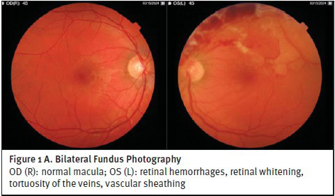
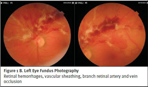
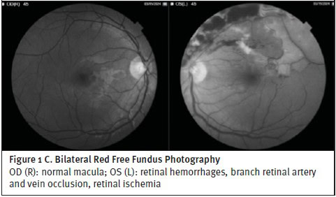
Ophthalmology referred the patient to the ED where he was admitted for further workup. Complete blood count (CBC), comprehensive metabolic panel (CMP), erythrocyte sedimentation rate (ESR) and C-reactive protein (CRP) were all within normal limits. Syphilis screen and blood cultures were negative. Computed tomography (CT) of the head, magnetic resonance imaging (MRI) of the head, computed tomography angiogram (CTA) of the head and neck, echocardiogram, and electrocardiogram were all normal. The patient was admitted for further evaluation. Hypercoagulability and autoimmune workups were performed but were pending at the time of discharge. In the hospital, he was given 1 dose of acetazolamide for concerns of elevated intraocular pressure, aspirin 324 mg for headache, and atorvastatin 40 mg for suspected ocular inflammation. The patient was discharged on aspirin 81 mg/day and valacyclovir 1 g/day for concerns of herpes simplex ophthalmicus.
The patient was seen in the ophthalmology clinic a few days later and stated that his vision was unchanged. Visual acuity and intraocular pressure were within normal limits, and fundoscopic imaging was unchanged since the previous visit. Repeat ophthalmological examination revealed 1–2+ cells in the anterior chamber and 2–3+ cells in the anterior vitreous.
A few days later, autoimmune serologies for HLA-B51 and -B54 were positive, so the patient was started on 40 mg of oral prednisone and referred to rheumatology. He returned a few days later and reported that vision in his left eye was greatly improved after taking the prednisone. Ophthalmologic exam showed no cells in the anterior chamber, 2-3+ cells still present in the anterior vitreous, and improvement of the centrally sparing retinal whitening. At this point, the patient was given a preliminary diagnosis of Behçet disease.
Discussion
Epidemiology
Behçet disease usually presents between the ages of 20–40 years. The prevalence and disease severity is higher in males in the Middle East and Mediterranean regions, and higher in females in North America, Northern Europe, and East Asia.4 The pathophysiology of the disease is not fully understood but shares some similarities with other autoimmune diseases given its autoinflammatory, autoimmune, and spondyloarthropathy-like features that can coexist.
Clinical Presentation
In most patients with Behçet disease (~95%), the primary presentation is recurrent oral aphthous ulcers on an erythematous base, located primarily on the lips, gingiva, cheeks, and tongue. Genital ulcers are primarily located on the scrotum or labia and are present in 65–75% of patients.[5] However, this patient only presented with very few small oral ulcers. Ocular manifestations can be found in ~50% of patients and generally include anterior, intermediate, posterior, or panuveitis with a relapsing-remitting course.5 Retinal vasculitis is seen in about half of patients with uveitis and can be complicated by retinal or vitreous hemorrhage, retinal ischemia, neovascularization, and secondary neovascular glaucoma. Macular edema, papillitis, and retinitis can also develop. Ocular involvement is the first sign of Behçet disease in about 20% of patients.5 Other extra-ocular manifestations of Behçet disease include: neurologic involvement (5%) presenting as parenchymal lesions; articular involvement (50–80%) presenting as recurrent inflammatory mono or oligoarthritis; gastrointestinal involvement presenting as diarrhea, hemorrhage, or perforation (4–38%); and cardiovascular involvement (2.2–50%) presenting as venous and arterial thrombosis and aneurysm.5 Gastrointestinal and cardiovascular involvement varies with geographical differences in prevalence.[6]
Workup
During the diagnostic workup, the patient in this case was found to be HLA-B51 positive, which has a high correlation with Behçet disease. The overall risk for developing Behçet disease is 5-10 times greater in patients who are HLA-B51 positive.[7] HLA-B51 positivity is seen in ~57% of patients globally with Behçet disease.7 However, HLA-B51 positivity is seen in only 20–30% of Behçet patients in North America.4 Other diagnostic tests to help rule in Behçet disease include the pathergy test. However, diagnosis varies heavily based on geographic location. In Middle Eastern patients with Behçet disease, 60% will test positive on a pathergy test, while only 5% of North American patients will have a positive result.6
Diagnostic Criteria
The diagnostic criteria for Behçet disease are based on clinical and laboratory findings. There are 3 accepted criteria: the International Criteria created in 2014,[8] the International Study Group Criteria created in 1990,[9] and the Japanese Criteria created in 1988.[10] While the International Study Group Criteria is the most widely used, the International Criteria is more sensitive. The patient in this case had a score of 4 points (2 points for oral ulcers and 2 points for ocular lesions). A score of at least 4 points is needed for diagnosis.8
| Oral ulcers | 2 points |
| Genital ulcers | 2 points |
| Ocular lesions | 2 points |
| Skin lesions | 1 point |
| Neurological manifestations | 1 point |
| Vascular manifestations | 1 point |
| Positive pathergy test | 1 point |
A score of at least 4 points is needed for diagnosis.
Case Conclusion
While the patient was a young male with HLA-B51 positivity, he lacked the oral and genital ulcers that are most often seen in Behçet patients. Ocular involvement is a major cause of morbidity in patients. It is the first sign of disease in ~20% of patients and is more frequent and more severe in males.4 Given this, clinicians must have a high degree of suspicion for Behçet disease with atypical presentations. Like most autoimmune conditions, Behçet disease responds well to initial high-dose steroids. As seen in this case, the patient’s condition stabilized after being on regular oral prednisone and his retinal vasculitis resolved. High-dose steroids can also be given along with azathioprine at the direction of the ophthalmology consult.[11]
Per the European Alliance of Associations for Rheumatology, monoclonal anti-TNF alpha antibodies, preferably infliximab, should be given in combination with other immunosuppressives. Glucocorticoids generally are not recommended as monotherapy. However, glucocorticoids help provide rapid control of inflammation, and the addition of immunosuppressives soon after is recommended.[12]
Bilateral involvement is the most common manifestation of ocular Behçet’s seen in ~80% of patients with ocular symptoms.4 However, in most patients with unilateral disease, bilateralization will usually occur within 2 years after the initial presentation.[13] Furthermore, about half of patients with uveitis will have coexisting retinal vasculitis as seen in this patient.4 Blindness will also occur in 10-20% of patients at 5 years with ocular involvement despite treatment.[14]
Disposition
Due to his sudden vision loss, the patient was referred to ophthalmology from urgent care. From there, the patient was referred to the ED for further evaluation. Initial CBC, CMP, ESR, and CRP were all within normal limits. Syphilis screen, blood cultures, CT head, MRI head, CTA head and neck, echocardiogram, and electrocardiogram were also negative. Based on his clinical symptoms and HLA-B51 positivity, he was diagnosed with Behçet disease.
Ethics Statement
The Helsinki Ethical Principles were met, and all criteria were followed regarding patient confidentiality and autonomy. The patient provided verbal consent for publication of this case.
Takeaway Points
- While eye complaints in urgent care are frequently non-emergent, providers should be aware of the potential presentation of Behçet disease.
- Behçet disease commonly presents in young men with recurrent oral and genital ulcers with possible systemic inflammation.
- One serious complication is monocular and binocular vision-threatening uveitis, which is the initial presentation in about 20% of patients.
- Initial management includes high-dose prednisone and urgent ophthalmology referral to avoid permanent vision loss.
Manuscript submitted September 23, 2025; accepted March 22, 2026.
References
- [1]. Vaziri K, Schwartz SG, Flynn HW Jr, Kishor KS, Moshfeghi AA. Eye-related Emergency Department Visits in the United States, 2010. Ophthalmology. 2016;123(4):917-919. doi:10.1016/j.ophtha.2015.10.032
- [2]. Mahjoub H, Ssekasanvu J, Yonekawa Y, et al. Most Common Ophthalmic Diagnoses in Eye Emergency Departments: A Multicenter Study. Am J Ophthalmol. 2023;254:36-43. doi:10.1016/j.ajo.2023.03.016
- [3]. Dag Y, Aydın S, Kumantas E. The profile of patients attending to the general emergency department with ocular complaints within the last year: is it a true ocular emergency?. BMC Ophthalmol. 2024;24(1):330. Published 2024 Aug 7. doi:10.1186/s12886-024-03608-1
- [4]. Emmi G, Bettiol A, Hatemi G, Prisco D. Behçet’s syndrome. Lancet. 2024;403(10431):1093-1108. doi:10.1016/S0140-6736(23)02629-6
- [5]. Davatchi F, Chams-Davatchi C, Shams H, et al. Behcet’s disease: epidemiology, clinical manifestations, and diagnosis. Expert Rev Clin Immunol. 2017;13(1):57-65. doi:10.1080/1744666X.2016.1205486
- [6]. Greco A, De Virgilio A, Ralli M, et al. Behçet’s disease: New insights into pathophysiology, clinical features and treatment options. Autoimmun Rev. 2018;17(6):567-575. doi:10.1016/j.autrev.2017.12.006
- [7]. Takeno M. The association of Behçet’s syndrome with HLA-B51 as understood in 2021. Curr Opin Rheumatol. 2022;34(1):4-9. doi:10.1097/BOR.0000000000000846
- [8]. International Team for the Revision of the International Criteria for Behçet’s Disease (ITR-ICBD). The International Criteria for Behçet’s Disease (ICBD): a collaborative study of 27 countries on the sensitivity and specificity of the new criteria. J Eur Acad Dermatol Venereol. 2014;28(3):338-347. doi:10.1111/jdv.12107
- [9]. Criteria for diagnosis of Behçet’s disease. International Study Group for Behçet’s Disease. Lancet. 1990;335(8697):1078-1080.
- [10]. Mizushima Y. Recent research into Behçet’s disease in Japan. Int J Tissue React. 1988;10(2):59-65.
- [11]. Çakar Özdal P. Behçet’s Uveitis: Current Diagnostic and Therapeutic Approach. Turk J Ophthalmol. 2020;50(3):169-182. doi:10.4274/tjo.galenos.2019.60308
- [12]. Hatemi G, Ramiro S, Ozguler Y, et al. EULAR recommendations for the management of Behçet’s syndrome: 2025 update. Ann Rheum Dis. Published online March 23, 2026. doi:10.1016/j.ard.2026.02.009
- [13]. Tugal-Tutkun I, Gupta V, Cunningham ET. Differential diagnosis of Behçet uveitis. Ocul Immunol Inflamm. 2013;21(5):337-350. doi:10.3109/09273948.2013.795228
- [14]. Gueudry J, Leclercq M, Saadoun D, Bodaghi B. Old and New Challenges in Uveitis Associated with Behçet’s Disease. J Clin Med. 2021;10(11):2318. Published 2021 May 26. doi:10.3390/jcm10112318