
Published on
Download the article PDF: Accidental Tongue Ring Ingestion Balancing Caution And Conservative Management In Urgent Care
Urgent Message: Most ingested foreign objects pass spontaneously with little risk of complications in adults. However, in cases where patients experience persistent symptoms, escalation to emergency department evaluation may be appropriate.
Naun Gutierrez Jr., MSN, FNP-C; Christina Gardner, DHSc, MBA, PA-C
Keywords: foreign body ingestion; gastrointestinal foreign body; tongue ring ingestion; radiography; conservative management; urgent care
Abstract
Introduction: Foreign body ingestion is a clinically important presentation in urgent care, with management guided by object characteristics, location in the gastrointestinal tract, and patient symptoms.
Presentation: A 21-year-old woman presented to urgent care 2 days after accidentally swallowing a tongue ring. She was tolerating oral intake without difficulty but reported mild persistent lower abdominal pain. She denied nausea, vomiting, hematemesis, melena, dysphagia, or bowel habit changes.
Physical Exam: Vital signs were within normal limits. She was well appearing and in no acute distress. Abdominal examination demonstrated mild tenderness to palpation in the lower quadrants without guarding or rebound. Cardiopulmonary and oropharyngeal examinations were unremarkable.
Diagnostic Evaluation and Diagnosis: Abdominal radiography demonstrated a linear-appearing metallic foreign body within the cecum measuring approximately 21 × 1 mm, without evidence of obstruction or perforation, consistent with ingested jewelry.
Management and Outcome: Given persistent abdominal tenderness in the setting of a distal gastrointestinal foreign body, the patient was referred to the emergency department (ED) for further evaluation. No additional diagnostic testing was required in the ED. She remained clinically stable during observation and was discharged home with return precautions.
Conclusion: This case highlights risk stratification and clinical decision-making in urgent care for distal gastrointestinal foreign bodies, emphasizing the role of symptom assessment, imaging, and determination of escalation of care.
Introduction
Foreign body ingestion occurs in both pediatric and adult populations.[1],[2] In adults, ingestion is most often accidental and commonly involves objects such as dental hardware, jewelry, or food-related materials.1,2 Most ingested foreign bodies pass through the gastrointestinal tract without intervention, with approximately 80–90% resolving spontaneously.[3],[4] However, clinical risk is highly dependent on object characteristics. Sharp objects, magnets, and button batteries are among those associated with increased risk of obstruction, perforation, or tissue injury, requiring urgent evaluation.[5],[6],[7] Initial evaluation includes symptom assessment and imaging to characterize the ingested object and guide clinical decision-making.4,5
Case Presentation
A 21-year-old woman with no significant medical history presented to an urgent care clinic 48 hours after accidentally swallowing her tongue ring. She did not seek immediate care, anticipating spontaneous passage of the object. She complained of mild but persistent lower abdominal pain that started the morning of presentation. She continued to eat and drink normally and denied nausea, vomiting, melena, hematochezia, hematemesis, swallowing difficulties, or any other bowel habit changes. She had no prior gastrointestinal disease or abdominal surgeries.
On examination, the patient’s vital signs were as follows: blood pressure of 123/78 mmHg; heart rate of 78 beats per minute; a respiratory rate of 17 breaths per minute; temperature of 37.1°C (98.9°F); oxygen saturation of 98% on room air; and a pain score of 2/10. She appeared well, alert, and in no acute distress. General examination was normal. Oral examination revealed moist mucous membranes without lesions, swelling, or visible foreign body. Oropharyngeal examination was otherwise normal. Cardiovascular examination demonstrated a regular rate and rhythm without murmurs, rubs, or gallops. Pulmonary examination revealed clear breath sounds bilaterally with no wheezes, rales, or rhonchi and no signs of respiratory distress. Abdominal examination revealed mild tenderness to palpation in the right and left lower quadrants without rebound tenderness, guarding, or rigidity.
Abdominal radiographs showed a linear-appearing metallic object in the cecum measuring approximately 21 × 1 mm. There was no evidence of obstruction, free air, or other acute findings. The radiologist recommended conservative outpatient management, expecting spontaneous passage within 48 hours. Despite the object’s low-risk characteristics and distal progression, the urgent care team elected to refer the patient to the emergency department (ED) because of persistent abdominal tenderness and the inability to provide prolonged observation within the urgent care setting.
In the ED, the patient underwent further clinical evaluation, during which she remained hemodynamically stable with no progression of symptoms. Urgent care radiographs, reviewed through the shared institutional imaging system, confirmed the location of the tongue ring in the cecum without evidence of obstruction, perforation, or other acute complication. Given the reassuring clinical presentation and imaging findings, no additional diagnostic testing was pursued. Her abdominal discomfort improved with conservative management during observation, and she continued to tolerate oral intake without difficulty. The patient was subsequently discharged home in stable condition with return precautions. She was scheduled for a follow-up appointment with her primary care provider the next day.
Differential Diagnosis and Medical Decision-Making
The differential diagnosis included gastrointestinal perforation, bowel obstruction, mucosal injury, and localized bowel irritation secondary to foreign body ingestion. Although imaging demonstrated the tongue ring within the cecum without evidence of obstruction or perforation, the patient’s persistent lower abdominal pain raised concern for possible early mucosal irritation or evolving complication. Stable vital signs and reassuring imaging findings reduced the likelihood of acute surgical pathology but did not fully exclude early complications.
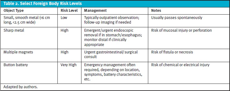
Key factors guiding management included the object’s size, smooth contour, and distal cecal location. The tongue ring measured less than 6 cm in length and 2.5 cm in width, classifying it as low risk and likely to pass spontaneously.5 However, given the presence of persistent abdominal pain and the limitations of urgent care monitoring, the decision was made to refer the patient to the emergency department for further evaluation and observation. This decision reflected a cautious, patient-centered approach that balanced low-risk object characteristics with clinical symptoms, incorporating inter-provider collaboration and adherence to evidence-based guidelines.4 After observation in the ED, repeat assessment showed no progression or peritoneal signs.
Final Diagnosis
The final diagnosis was accidental ingestion of a tongue ring, located in the cecum, in an otherwise healthy adult without acute complications.
Patient Perspective
The patient appreciated the detailed explanation and rationale for the referral to the ED. She understood the conservative approach and felt reassured by clear ED discharge instructions for home monitoring and follow-up. She reported confidence in safely tracking the object’s passage and acknowledged the benefits of interdisciplinary collaboration.
Discussion
National data estimate there were more than 900,000 foreign body ingestion cases during 2014–2023 in the United States, with increasing incidence among adults.[8] Approximately 90% of patients are discharged after evaluation without requiring admission or procedural intervention.1,3,4 However, adult cases are more frequently associated with hospitalization and higher-risk ingestion patterns, particularly in populations with underlying comorbidities or intentional ingestion.[9]
Clinical presentation depends on object type, size, and anatomic location. Many patients with uncomplicated foreign body passage to the distal gastrointestinal tract remain asymptomatic; however, symptoms such as abdominal pain, vomiting, drooling, chest discomfort, or signs of peritonitis may indicate complications and warrant further evaluation.3 In this case, the patient’s persistent abdominal pain raised concern for possible localized irritation or early complication, prompting closer clinical assessment. Although the tongue ring had progressed to the cecum and possessed low-risk characteristics (small size, blunt contour, and distal location), the presence of persistent abdominal pain complicated an otherwise straightforward conservative management pathway.
Current guidelines generally support outpatient observation for asymptomatic patients with blunt foreign bodies that have passed beyond the stomach (Tables 1–2).4 However, persistent abdominal symptoms warrant careful reassessment because early perforation, localized inflammation, or evolving obstruction may initially present with subtle findings despite reassuring imaging.3 In this case, escalation to the ED was primarily driven by persistent abdominal pain despite distal progression of the object. The referral reflected a cautious approach intended to facilitate further monitoring and rapid access to advanced imaging or specialty consultation if symptoms progressed.4
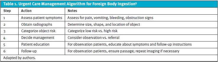
Diagnostic evaluation begins with a focused history and physical examination. Plain radiography is typically recommended for suspected radiopaque objects to determine the presence, size, number, and location of the foreign body.4,5 Computed tomography should be considered when there is concern for perforation or other complications.4 In this case, imaging confirmed the object’s location within the cecum and supported risk stratification.
Management is time-sensitive and depends on the object’s characteristics, location, and the patient’s clinical status. Emergent endoscopy (preferably within 2 hours and no later than 6 hours) is indicated for complete esophageal obstruction and for sharp-pointed objects or batteries in the esophagus. Other high-risk objects, including magnets, may require urgent intervention depending on their number and location.4,7 Urgent or nonurgent endoscopy may be appropriate for other retained objects depending on size and progression. Objects that are long (greater than 5–6 cm) or wide (greater than 2–2.5 cm) are less likely to pass spontaneously and may require intervention.4,[5] In contrast, asymptomatic patients with small, blunt objects that have progressed beyond the stomach can often be managed conservatively with observation alone.
Special scenarios require additional consideration. Button battery ingestion requires prompt evaluation and management based on the battery’s location and clinical circumstances, and ingestion of multiple magnets poses a risk of bowel necrosis from pressure effects. Concealed packets of drugs that have been swallowed (“body packing”) are typically managed with close observation rather than endoscopic retrieval. Surgical intervention is reserved for suspected obstruction or packet rupture.4 Food bolus impaction may require endoscopic management and further evaluation for underlying esophageal pathology, such as eosinophilic esophagitis.[11]
Ethics Statement
Patient consent for publication was obtained in accordance with institutional guidelines. No identifiable patient information is included in this case report.
Takeaway Points
- Foreign body ingestion in adults is most often low risk, as most objects without high-risk features pass spontaneously once beyond the stomach.
- Clinical decision-making in urgent care relies on integrating symptom assessment, imaging findings, and object characteristics to appropriately stratify risk.
- Persistent abdominal symptoms despite distal progression of a foreign body warrant careful reassessment and may justify escalation of care for further evaluation.
- High-risk ingestions, including button batteries, sharp objects, and magnets, require prompt recognition due to increased risk of severe complications.
- Patient education, return precautions, and inter-provider communication are essential components of safe, evidence-based management of foreign body ingestion.
Manuscript submitted January 6, 2026; accepted August 14, 2026.
References
- [1]. Hsieh A, Hsiehchen D, Layne S, Ginsberg GG, Keo T. Trends and clinical features of intentional and accidental adult foreign body ingestions in the United States, 2000 to 2017. Gastrointest Endosc. 2020;91(2):350-357.e1. doi:10.1016/j.gie.2019.09.010.
- [2]. Orsagh-Yentis D, McAdams RJ, Roberts KJ, McKenzie LB. Foreign-body ingestions of young children treated in US emergency departments: 1995–2015. Pediatrics. 2019;143(5):e20181988. doi:10.1542/peds.2018-1988.
- [3]. Ambe P, Weber SA, Schauer M, Knoefel WT. Swallowed foreign bodies in adults. Dtsch Arztebl Int. 2012;109(50):869-875. doi:10.3238/arztebl.2012.0869.
- [4]. Birk M, Bauerfeind P, Deprez PH, et al. Removal of foreign bodies in the upper gastrointestinal tract in adults: European Society of Gastrointestinal Endoscopy (ESGE) clinical guideline. Endoscopy. 2016;48(5):489-496. doi:10.1055/s-0042-100456.
- [5]. Lee SM, Baek SE, Lee CW, Kim YC, Kim MJ. Foreign Body Ingestion: Radiologic Evaluation, Findings, and Management. Korean J Radiol. 2025;26(7):638-649. doi:10.3348/kjr.2025.0118
- [6]. Kinjalk M, Sehgal M, Ratan SK, et al. Foreign body ingestion in children: an experience of 99 cases in a tertiary care center in Delhi. J Indian Assoc Pediatr Surg. 2024;29(3):223-232. doi:10.4103/jiaps.jiaps_183_23.
- [7]. Chirica M, Kelly MD, Siboni S, et al. Esophageal emergencies: WSES guidelines. World J Emerg Surg. 2019;14:26. doi:10.1186/s13017-019-0245-2.
- [8]. Lad H, Durant S, Yan K, Kaye R. Trends and outcomes of foreign-body ingestion: national emergency department data over a decade. Laryngoscope. 2026;136(7):2938-2946. doi:10.1002/lary.70477.
- [9]. Acehan S, Satar S, Gulen M, et al. Foreign body ingestion in adults: clinical challenges and outcomes in high-risk populations presenting to the emergency department. Eur J Gastroenterol Hepatol. 2026;38(5):546-553. doi:10.1097/MEG.0000000000003106.
- [10]. Sbeit W, Basheer M, Shahin A, Makanovitsky D, Khoury T. Predictors of spontaneous passage of ingested foreign bodies in adults: twelve years of experience. Surg Endosc. 2024;38(3):1533-1540. doi:10.1007/s00464-023-10657-y.
- [11]. Kamalumpundi V, Saha B, Leggett C. Management of foreign body ingestions and food impactions: advice for general practitioners. Mayo Clin Proc. 2025;100(8):1411-1419. doi:10.1016/j.mayocp.2025.03.035.
Author Affiliations: Naun Gutierrez Jr., MSN, FNP-C, Carilion Clinic, Roanoke, Virginia. Christina Gardner, DHSc, MBA, PA-C, Carilion Clinic, Roanoke, Virginia; Jefferson College of Health Sciences, Roanoke, Virginia. Authors have no relevant financial relationships with any ineligible companies.
