Published on
Differential Diagnosis
- Diffuse panbronchiolitis
- Pulmonary metastases
- Septic pulmonary emboli
- Silicosis
- Tuberculosis
Diagnosis
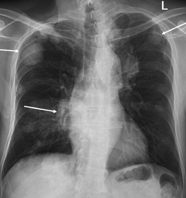
The x-ray shows multiple bilateral, well-circumscribed, rounded masses. The largest appears in the right posterior perihilar region. There are many causes for multiple pulmonary masses, however metastases are the most common.
Learnings and What to Look for
- The priority is to identify the lesion in the lung and distinguish a nodule from a mass
- A pulmonary nodule is defined as a small (<30 mm), well-circumscribed lesion, completely surrounded by normal lung parenchyma
- A pulmonary mass is defined as a pulmonary opacification of >30 mm
- Common causes of pulmonary nodules are hamartomas, mucous gland adenoma, histiocytoma, and lipomas. The most common cause of pulmonary mass is malignancy. Other causes of pulmonary masses include autoimmune disease, fungal infections, and tuberculosis.
Pearls for Urgent Care Management
- After ruling out cardiac causes for the patient’s symptoms, additional imaging with a CT scan is indicated
- A rapid referral to an oncologist is often frequently warranted
Acknowledgment: Image and case provided by Experity Teleradiology (www.experityhealth.com/teleradiology).
A 75-Year-Old with Chest Pressure
1 2