Published on
Differential Diagnosis
- Epidermoid cyst
- Calcinosis cutis
- Cutaneous tuberculosis
- Furunculosis
Diagnosis
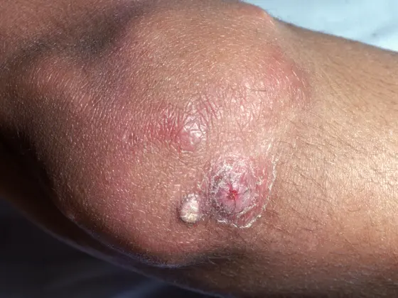
The correct diagnosis is calcinosis cutis, or cutaneous calcification—the deposition of insoluble calcium salts in the skin and subcutaneous tissue due to local dysregulation of calcium metabolism. In children, there is a male predilection with an earlier age of onset. Dystrophic calcification is typically seen in autoimmune connective tissue diseases, most commonly in juvenile dermatomyositis (DM) and the CREST form of systemic sclerosis, and usually arises in damaged skin in which local calcium metabolism is altered, allowing intracellular crystallization in the setting of normal serum calcium and phosphorus levels
Learnings/What to Look for
- The skin lesions are typically crusted, firm and jagged nodules from chalk-like calcium that can extrude through the skin, which may be painful or susceptible to secondary infection
- Approximately 50%-70% of children with juvenile DM will develop calcinosis cutis or some form of cutaneous calcification, in contrast to 10%-20% of patients with adult DM
- The most frequently affected sites in DM are the elbows, knees, buttocks, and shoulders
- In the CREST form of systemic sclerosis, the hands and upper extremities, often over bony prominences and tendons, are most commonly affected
Pearls for Urgent Care Management
- In the case of a child, advise the caregiver that the patient should minimize cold exposure and situations that may result in local trauma
- Depending on the size and severity of the lesions, diltiazem, bisphosphonates, probenecid, aluminum hydroxide, warfarin, ceftriaxone, and intravenous immunoglobulin have been found to be successful in treating calcinosis cutis
- Surgical excision and carbon dioxide laser can also be used
Acknowledgement: Image and case provided by VisualDx (www.VisualDx.com/jucm).
Download the article PDF: A 7-Year-Old Male with Lesions on His Knees