Published on
Urgent message: Epiglottitis is classically viewed as a pediatric disease, but has become increasingly common in the adult population. While symptoms may present as an isolated sore throat, they can quickly progress to complete airway compromise with need for emergency cricothyroidotomy. Due to the high risk for morbidity and mortality, urgent care providers must maintain a high index of suspicion to avoid misdiagnosing a potentially catastrophic disease.
Zachary DePriest, MS, PA-C
INTRODUCTION
Adult epiglottitis (AE) is a potentially life-threatening condition. While historically thought to be a disease of childhood, advent of the Haemophilus influenza B (HiB) vaccine in the 1980s has reduced mortality to 7% (consequently increasing relative incidence among adults).1
CASE PRESENTATION
A 69-year-old female with a history of hypertension and dyslipidemia presented with a 3-day history of increasing pharyngitis, odynophagia, mild shortness of breath, subjective fever, and progressive hoarseness. She denied any drooling or inability to handle secretions, although she did note that her discomfort was greatest on the right side of her throat. On exam, the patient was febrile at 38⁰C but the rest of her vital signs were normal aside from a mild tachycardia at 106 bpm. Blood pressure was 130/78 mmHg, respiratory rate of 18 breaths/minute, and oxygen saturation of 98% on room air. She was noted to be moderately hoarse but handling secretions without difficulty or stridor. The posterior oropharynx was mildly erythematous but without edema, exudate, or asymmetry. There was bilateral submandibular and anterior cervical lymphadenopathy most pronounced on the right. Heart rate was regular with normal heart tones and breath sounds were clear with good aeration. She had normal mentation.
Differential Diagnosis
- Group A beta-hemolytic streptococcal pharyngitis (GABHS)
- Viral pharyngitis
- Laryngitis
- Peritonsillar abscess
- Epiglottitis
- Retropharyngeal abscess
- Ludwig’s angina
- Lemierre’s syndrome
Diagnostics
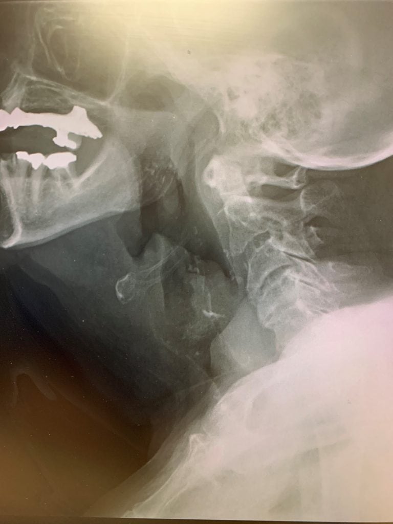
Due to the progressive hoarseness with relatively normal physical exam findings, consideration was given to deep space infection and the patient was sent for a lateral soft tissue neck x-ray. This demonstrated diffuse supraglottic edema and a prominent epiglottis for which the radiologist recommended CT soft tissue neck with IV contrast (Figure 1).
Course and Treatment
The facility this patient presented to was a standalone urgent care center (UCC) with laboratory, ultrasound, and x-ray capability. There was no CT scanner on site. It was recommended to transport the patient via EMS with an Advanced Life Support (ALS) crew capable of managing the patient’s airway if it became compromised. The patient refused transport, insisting upon driving herself to the ED. She was given 10 mg PO dexamethasone prior to discharge. The patient arrived in the ED a short time later, where she underwent additional workup that revealed a leukocytosis of 18,000 with normal renal function and electrolytes. She was given IV fluids and 1 g ceftriaxone while her airway was monitored. Contrast-enhanced CT of the neck revealed:
- Markedly edematous and irregularly enhancing soft tissues of the epiglottis and right pharyngeal wall extending into the true vocal cords. A moderate degree of fat stranding was appreciated, suggesting epiglottitis; however, malignancy cannot be excluded.
- Moderate glottic airway narrowing.
The patient underwent nasopharyngoscopy in the ED by the ENT specialist, revealing an inflamed and edematous epiglottis consistent with epiglottitis.
RESOLUTION OF CASE
The patient was admitted to the ICU for airway monitoring and continued dexamethasone and ceftriaxone. The next day she noted significant improvement in her discomfort with improved phonation. Repeat nasopharyngoscopy the following day revealed nearly resolved epiglottic edema and the patient was discharged on day 5 to finish a 14-day course of amoxicillin clavulanate.
DISCUSSION
Sore throat is a common complaint in the urgent care setting in both the adult and pediatric populations, accounting for over 11% of visits overall.2 The vast majority of cases are viral in etiology (40%-60%) due to rhinoviruses, influenza A and B, parainfluenza viruses, Epstein-Barr virus (infectious mononucleosis), and adenovirus. These require only supportive management.3 GABHS pharyngitis accounts for only 5%-15% of all adult cases of pharyngitis and 15% to 36%in children but has significant nonsuppurative (rheumatic fever, glomerulonephritis) and suppurative complications (peritonsillar abscess).4 Other less-frequent bacterial etiologies include Group C and D strep, Fusobacterium necrophorum (Lemierre’s syndrome), Mycoplasma pneumoniae, Neisseria gonorrhea, and Corynebacterium diphtheriae (diphtheria).5
Epiglottitis is an acute inflammation of the epiglottis and supraglottic structures that can lead to acute airway obstruction. Haemophilus influenza used to be the most common pathogen prior to development of the HiB vaccine but is now increasingly caused by Streptococcus pneumoniae, Staphylococcus aureus, and GABH.6,7 Due to immunization, epiglottitis has decreased in the pediatric population and is now increasingly seen in adults, most notably between the ages of 45 and 64.8 Prior to the 1980s, the child:adult ratio of epiglottitis was 2.6:1. By the mid 1990s, that ratio had reversed to 0.4:1 (child:adult).9 Consider the “3 Ds” in the pediatric population:
- Drooling
- Dysphagia
- Distress
Adults with epiglottitis, however, tend to present with more subacute and insidious complaints that include pharyngitis, odynophagia, and fever.10 Red flags to always evaluate for are changes in phonation (hoarseness or muffled voice), stridor, tripod position, and inability to handle secretions. Toxic or super-heated inhalations such as with crack cocaine use can also cause noninfectious epiglottitis. The clinician should consider imaging in any patient who appears ill but has an unremarkable oropharynx or with voice changes such as hoarseness or a “hot potato voice.” Lateral soft tissue neck x-ray is available in most UCCs and carries a 90% sensitivity with the classic finding being a “thumbprint” sign indicative of an edematous epiglottis. The ratio of the width of the epiglottis to the anteroposterior width of C4 should not exceed 0.33 (sensitivity 96%, specificity 100%).11,12 Surprisingly, there is no described sensitivity or specificity in the literature for CT neck with IV contrast.13 This imaging modality is primarily used to differentiate other suppurative conditions such as PTA or on equivocal plain films.14 One must use CT with caution, however, as it typically involves significant time in the radiology department away from definitive airway management.The gold standard of diagnosis is direct laryngoscopy.
Airway management is the main priority. Supplemental oxygen should be applied with difficult airway cart, fiber optic bronchoscopy, and cricothyrotomy kit at the bedside. Broad-spectrum antibiotics are indicated in the form of a third-generation cephalosporin such as ceftriaxone 2g IV daily or ampicillin/sulbactam 3 g q6h. Vancomycin 15 mg/kg should be added q12h in the critically ill patient or if there is clinical concern for methicillin-resistant Staphylococcus aureus (MRSA) infection. Corticosteroids such as dexamethasone 10 mg IV are frequently used; however, their efficacy is somewhat controversial.15
All patients with epiglottitis need to be admitted to a monitored bed with continuous airway monitoring, preferably in an ICU setting.
SUMMARY
- Epiglottitis is increasingly seen in the adult population due to advent of the Hib vaccine.
- Beware of voice changes; it is reasonable to obtain imaging studies in a patient who reports a change in phonation or in whom you notice a muffled voice.
- Epiglottitis is a true airway emergency.
- Stridor in a patient with epiglottitis is indicative of impending airway collapse and need for immediate intubation or cricothyrotomy.
- Any patient in whom you suspect epiglottitis needs to be emergently transported to a facility where definitive airway management can be accomplished by an ENT, anesthesia, or a surgical specialist.
REFERENCES
- Ames WA. Adult epiglottitis: an under-recognized, life-threatening condition. Br J Anaesth. 2000;85(5):795-797.
- The Journal of Urgent Care Medicine. 2018 Urgent Care Chart Survey.
- Santos M, Smith M, Effron D. Epiglottitis: old problem, new patients. Emerg Phys Monthly. Available at: http://epmonthly.com/article/epiglottitis-old-problem-new-patients/. Accessed October 5, 2020.
- Wilson A. Pharyngitis. In: Skolnik NS, ed. Essential Infectious Disease Topics for Primary Care. New York, NY: Springer;2008: 15-24.
- Bisno AL. Acute pharyngitis: etiology and diagnosis. Pediatrics. 1996;97(6):949-954.
- ACEP Now. August 1, 2010. Sore Throats—What Really Works? https://www.acepnow.com/article/sore-throats-really-works/. Accessed October 2, 2020.
- Angirekula V, Multani A. Epiglottitis in an adult. N Engl J Med. 2015;372:e20
- Shah RK, Stocks C. Epiglottitis in the United States: national trends, variances, prognosis, and management. Laryngoscope. 2010;120(6):1256–1262.
- Berg S, Trollfors B, Nylen O, et al. Incidence, aetiology, and prognosis of acute epiglottitis in children and adults in Sweden. Scand J Infect Dis. 1996;28(3):261-264.
- Alcaide ML, Bisno AL. Pharyngitis and epiglottitis. Infect Dis Clin North Am. 2007;21(2):449-469.
- Nemzek W. A reappraisal of the radiologic findings of acute inflammation of the epiglottis and supraglottic structures in adults. Am J Neuroradiol. 1995;16:495-502.
- Rothrock SG. Radiologic diagnosis of epiglottitis: objective criteria for all ages. Ann Emerg Med. 1990;19(9):978-982.
- Ramalanjaona G, Shpak M. Challenges in diagnosing adult epiglottitis: limitations of CT scan. Emerg Med Open Journal. 2014;1(1):1-4.
- Smith M. CT in adult supraglottitis. Am J Neuroradiol. 17:1355-1358.
- Cirilli AR. Emergency evaluation and management of the sore throat. Emerg Med Clin North Am. 2013;31(2):501-515.
Zachary DePriest, MS, PA-C is Director of Education and Staff Development, Alteon Health Midwest. The author has no relevant financial relationships with any commercial interests.