Published on
What Does This ECG Show?
- Acute inferior STEMI
- Left axis deviation (LAD)
- Left bundle branch block
- Left ventricular hypertrophy
- Paced rhythm
Diagnosis
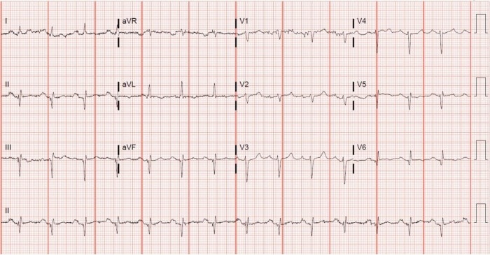
The ECG reveals a sinus rhythm at a rate of 84 beats per minute. There is left axis deviation, first degree AV block, and a narrow QRS and normal QT interval. There are no signs of acute ischemia. The diagnosis is left axis deviation (LAD).
Learnings/What to Look for
The QRS axis describes the direction of the vector of ventricular depolarization and normally lies between -30° and +90° or generally in the direction of lead II (+60°). The QRS axis can be estimated using a quadrant method with lead I and aVF as the x and y axes respectively. Begin by determining the predominant direction of the QRS complex in leads I and aVF (i.e. mostly positive or mostly negative).
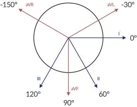
For example, along the x axis (using lead I), a mostly positive QRS complex points rightward. Along the y axis (using lead aVF), a mostly positive QRS complex points downward. Together, the combined forces point to the quadrant between 0° and +90°.
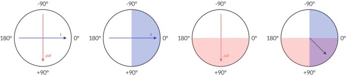
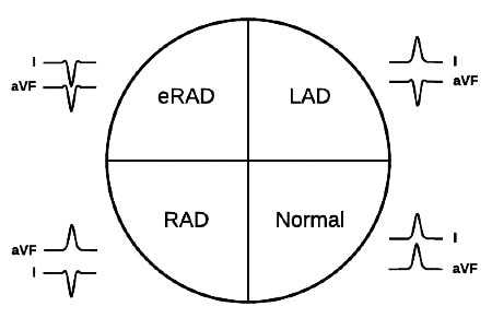
Our patient has a mostly positive QRS in lead I and a mostly negative QRS in lead aVF. The resultant vector points up and to the right or left axis deviation. Each quadrant is named (see Figure 3) and has a differential diagnosis of possible etiologies. The possible causes of left axis deviation include:
- Left ventricular hypertrophy
- Left bundle branch block
- Paced rhythm
- Ventricular ectopy
- Left anterior fascicular block
- Ventricular pre-excitation
7. Prior inferior myocardial infarction (inferior Q waves)
Left axis deviation may be caused by more depolarizing myocardium drifting forces further leftward as in left ventricular hypertrophy. Mechanical processes altering the heart’s position such as pregnancy or ascites can similarly deflect forces leftward. Conduction abnormalities interrupting the propagation of ventricular contraction along the normal axis including left bundle branch block, left anterior fascicular block and preexcitation syndromes (like Wok-Parkinson-White) can cause left axis deviation. Finally, a loss of viable myocardium, particularly involving the inferior wall, is a cause of left axis deviation. Our patient’s ECG shows large Q waves in the inferior leads (II, III, aVF), indicative of prior inferior myocardial infarction, causing a left axis deviation.
Pearls for Urgent Care Management
- The determination of QRS axis is a critical component of the systematic approach to ECG interpretation.
- An abnormal ECG axis is not independently pathologic but should prompt a hunt for the cause.
- Common causes of left axis deviation include:
- Left ventricular hypertrophy
- Blocks: left bundle branch block, left anterior fascicular block
- Ventricular pre-excitation
- Prior inferior myocardial infarction
References
- Kashou AH, Basit H, Chhabra L. Electrical right and left axis deviation. [Updated 2021 Jan 24]. StatPearls. January 2021. Available at: https://www.ncbi.nlm.nih.gov/books/NBK470532/. Accessed August 3, 2021.