
Published on
Download the article PDF: Clinical Image Challenges June 2026
Differential Diagnosis
- Slow atrial fibrillation
- ST-elevation myocardial infarction
- Hypercalcemia
- Osborn waves of hypothermia
- Junctional escape
Diagnosis
The diagnosis in this case is sinus bradycardia with Osborn waves of hypothermia. The ECG reveals sinus bradycardia with a ventricular rate of 48 beats per minute. Prominent J-point elevations immediately following the QRS complexes are present, consistent with Osborn waves associated with hypothermia.
Discussion
Electrocardiographic manifestations of hypothermia have been recognized for over a century and include sinoatrial exit block, PR and QT interval prolongation, QRS widening, ST-segment depression, and atrial and ventricular fibrillation. Osborn waves—upright J-point deflections immediately following the QRS complex—are more specific findings (Figure 2).1,2 These deflections may mimic ST-segment elevation and create the appearance of ST-segment concavity.
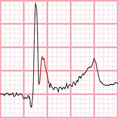
The electrocardiographic manifestations of hypothermia follow a predictable progression as core body temperature drops. These changes are fundamentally driven by the temperature-dependent slowing of myocardial conduction and repolarization. With mild hypothermia (32°–35°C), sinus bradycardia, shiver artifact (rapid, erratic, fine-to-coarse spikes that distort the isoelectric baseline), and interval prolongation (PR, QRS, and QT) are common. With moderate hypothermia (28°–32°C), sinus bradycardia frequently degenerates into atrial fibrillation, usually with a slow ventricular response. Osborn waves become distinctly visible and prominent, and shiver artifact often resolves. Severe hypothermia (< 28°C) is associated with ventricular dysrhythmia, high-grade atrioventricular blocks, and asystole.3
Although hypercalcemia can shorten the QT interval and produce J-point abnormalities that mimic Osborn waves, this ECG demonstrates distinct repolarization morphology clearly separate from the Osborn deflections.
This patient’s core temperature was 27°C (81°F), and he was immediately transferred to an emergency department for further treatment.
What to Look For
- Hypothermia manifests electrocardiographically via a predictable progression that includes sinus bradycardia, prolonged intervals, Osborn waves, slow atrial fibrillation, and ultimately degenerates into ventricular dysrhythmias and asystole.
- Osborn waves are upright J point deflections that immediately follow the QRS complex.
Pearls For Initial Management, Considerations For Transfer
- Passive and active rewarming measures are the mainstays of treatment.
- Urgent care centers are not generally equipped to treat hypothermia severe enough to produce ECG findings; immediate transfer is warranted.
- Warm blankets and/or forced-air warming devices may be initiated while awaiting transfer.
References
- Doshi HH, Giudici MC. The EKG in hypothermia and hyperthermia. J Electrocardiol. 2015;48(2):203-209. doi:10.1016/j.jelectrocard.2014.12.001
- Vassallo SU, Delaney KA, Hoffman RS, Slater W, Goldfrank LR. A prospective evaluation of the electrocardiographic manifestations of hypothermia. Acad Emerg Med. 1999;6(11):1121-1126. doi:10.1111/J.1553-2712.1999.TB00114.X
- Okumura H, Okada N, Hamanaka K, Okada Y, Kitamura T, Matsuyama T. Electrocardiographic patterns of accidental hypothermia. Am J Emerg Med. 2025;90:210-213. doi:10.1016/j.ajem.2025.01.079