Published on
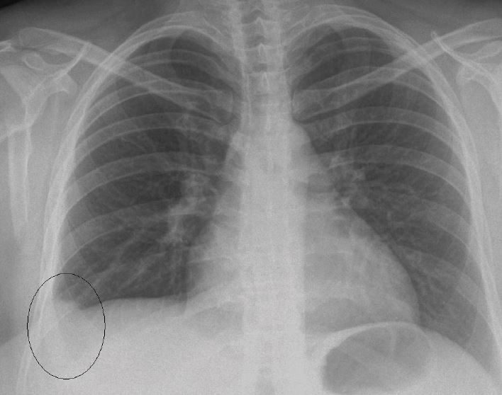
Initially, the radio-opacity seen in the right base was interpreted as pleural effusion. The official read of the chest x-ray led to suspicion of Hampton’s hump in the right lower lobe.
Though the patient never had any shortness of breath, in view of her unusual pain, pathological x-ray, recent childbirth, and obesity, she was referred to hospital, where chest computed tomography showed a massive pulmonary embolus (PE).
Conclusion
It was imperative to rule out PE in this case. Factors that might have led the physician to discount that possibility—no shortness of breath or signs of deep-vein thrombosis and an x-ray that failed to inspire suspicion—should be overshadowed by the patient’s risk factors and recognition that plain film may show little evidence of PE (Figure 2).