Published on
Physical Examination
On physical examination, the patient’s vital signs were as follows:
- Temperature: afebrile
- Pulse: 106 beats/min
- Respiration: 24 breaths/min
- Blood pressure: 138/92 mm Hg
The patient is alert and oriented. He winces when he sits forward for the lung examination and when taking a breath. He has slightly diminished lung sounds bilaterally, and no wheezing. His heart has a regular rate and rhythm without murmur, rub, or gallop. His abdomen is soft and nontender without rigidity, rebound, or guarding.
Urgent Care Work-Up
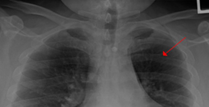
A chest x-ray was ordered, which showed a left posterior fourth-rib fracture.
Differential Diagnoses
- Pneumothorax
- Pulmonary infiltrate
- Pulmonary embolism
- Osteolytic lesion
- Flail chest
Diagnosis
The patient has a left posterior fracture of the fourth rib.
Learnings
A blow to the chest may cause such injuries as a contusion of the skin and chest wall, fractures, or injury to the lungs or great vessels. Rib fractures are the most common thoracic injury, present in 10% of all traumatic injuries. The fifth to ninth ribs are typically affected. A great amount of force is necessary to fracture the first and second ribs, so if this injury is present, serious intrathoracic injuries are more likely. Acute complications of rib fractures and underlying injuries include the following:
- Pneumothorax
- Hemothorax
- Lung contusion
- Injury to the great vessels
- Flail chest
- Injuries to adjacent structures such as intra-abdominal or neck injuries
- Sternal fracture
What To Look For
The medical history and physical examination should include exploration of the mechanism of injury: a fall, motor vehicle collision, assault, syncope, or abuse. Localize the area of greatest pain and inquire about what makes the pain worse, such as movement, breathing, or palpation. Assess for swelling and check the integrity of the skin. Evaluate for a seat belt sign if the patient was in a motor vehicle collision. Palpate for the area of greatest pain, because associated findings such as crepitus may indicate a pneumothorax. Look for secondary signs. Distended neck veins, tachycardia, tachypnea, and hypotension may indicate a tension pneumothorax or pericardial tamponade.
Imaging should include a posteroanterior and left lateral chest x-ray. A rib series was traditionally be ordered, but recent studies as well as guidelines from the American College of Radiology suggest that this is not necessary, because findings on such a series rarely change management. Specifically look for complications of rib fractures, including a pulmonary contusion, pneumothorax, or great vessel injury, including a traumatic aortic dissection.
Management in the urgent care includes encouraging the patient to perform coughing and deep-breathing exercises to keep the lungs expanded. Ambulation and activities of daily living should be continued, because bed rest may contribute to a rapid decline, particularly in the elderly. Pain control should include either hydrocodone-acetaminophen (Vicodin) or oxycodone-acetaminophen (Percocet) and not merely a codeine-containing product. Pain control is important so that the patient can aggressively pursue pulmonary hygiene. Additional analgesia can be obtained by adding anti-inflammatory medications such as ibuprofen (600 mg three times a day). This can be safely added to the narcotic medication, because nonsteroidal anti-inflammatory drugs are excreted by the kidneys.
Follow-up instructions should inform the patient of the consideration of pneumonia, although one recent study of 1057 patients demonstrated that such information was provided to only 0.5% of patients and was more likely to be provided to patients with a history of chronic obstructive pulmonary disease or asthma. Instruct patients to return if they develop fever, shortness of breath, increased pain, or dizziness.

