Published on

Physical Examination
On physical examination, the patient’s vital signs are as follows:
- Temperature: The patient is afebrile.
- Pulse: 104 beats/min
- Respirations: 20 breaths/min
- Blood pressure: 148/78 mm Hg
The patient is alert and oriented. He is not in acute distress, but he occasionally grimaces. His lungs are clear to auscultation, and he has a regular heart rate and rhythm without murmur, rub, or gallop. His abdomen is soft and nontender without rigidity, rebound, or guarding; it has no bruising; and it is not distended. He experiences pain with palpation of the right scapula, but he has no cuts or breaks in the skin there. The shoulder does have good range of motion, but he has significant pain with shoulder motion. Sensation over the distribution of the axillary nerve is intact grossly, and the radial pulse is 2+. There is no bruising of the skin over the shoulder.
Differential Diagnosis
- Anterior shoulder dislocation
- Posterior shoulder dislocation
- Scapular fracture
- Acromioclavicular separation
- Coracoid process fracture
- Acromion fracture
Urgent Care Workup
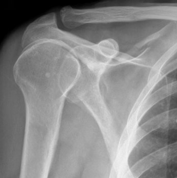
A radiograph (Figure 2) shows a fracture line through the superior scapular spine. There is no involvement of the glenoid or coracoid process.
Diagnosis
The patient has a scapular fracture.
Learnings
Motor vehicle accidents account for 50% of scapular fractures. The most common site of fracture is the body of the scapula or the glenoid neck. Scapular fractures are often associated with other injuries, such as pneumothorax and fractures of ribs, the sternum, and the clavicle.
The scapula is shaped like a triangle. It is evident on a shoulder x-ray as well as partially visible on a chest x-ray. The scapula stabilizes the upper extremity. It is a sturdy bone that requires a significant amount of force to fracture. It articulates with the humeral head at the glenoid, which can also be a fracture site.
What to Look For
A scapular fracture typically results from a significant mechanism of injury, such as a forceful fall onto the outstretched hand, directly on the shoulder, or a motor vehicle accident. When obtaining the medical history, ask about the mechanism of injury, location of pain, exacerbators of pain (such as movement through the range of motion), paresthesias, and other injuries, such as of the head or neck. A scapular fracture typically occurs during a high-energy injury, so pay close attention to the vital signs, particularly tachycardia, tachypnea, or hypotension, because such conditions may indicate injuries such as pulmonary contusion or pneumothorax in intrathoracic structures.
Physical examination will reveal that the patient is significant pain. Tachycardia may be present because of the pain or an associated injury. The patient will typically hold the arm in adduction. Document the range of motion for the shoulder, neck tenderness, visual evidence of head trauma, shoulder and elbow pain, and the neurovascular status, including pulses.
Testing initially involves obtaining a plain x-ray series. A scapular fracture can be seen on the anteroposterior view or on the lateral view of the shoulder, but a dedicated scapular series is preferred. Trace the cortex, looking for disruption, and examine trabeculations for lucency. The surrounding structures should be closely inspected because there may be a fracture of the ribs, clavicle, or humerus. Computed tomography will often have to be performed to define the extent of the injury; this can be completed in an emergency department.
In an urgent care center, initial management involves provision of adequate analgesia, immobilization, and radiographic imaging. The arm should be placed in a sling and swathe for comfort, because most scapular fractures are managed without surgery unless there is articular involvement of the glenoid or significant displacement. The patient should be referred to an orthopedic surgeon for follow-up. If there is hemodynamic instability, place an intravenous line, initiate fluid infusion, and arrange to transfer the patient to an emergency department.
With localized pain and a defined mechanism of injury, scapular fractures can be immobilized and the treatment can be handled on an outpatient basis, but maintain a high index of suspicion for associated injuries. If there is any concern whatsoever that there are associated life-threatening injuries, transfer the patient to an emergency department for further imaging, observation, and testing.
Key Points
- Scapular fractures typically occur because of a high-impact injury.
- The patient will report pain over the scapula, and this will be confirmed by physical examination.
- Maintain a high index of suspicion for associated injuries such as rib fractures, vascular injury, head injury, cervical fracture, sternal fracture, pulmonary contusion, and pneumothorax.
- Isolated scapular fractures are managed without surgery.