Published on
Differential Diagnoses
• Fungal keratitis
• Acanthamoeba keratitis
• Corneal foreign body
• Herpes simplex virus keratitis
Learnings
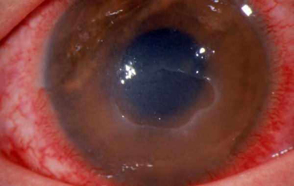
A bacterial corneal ulcer, or bacterial keratitis, is an infection of the corneal stroma that causes rapid vision loss and pain. Infectious corneal ulcers must be treated as soon as possible to preserve vision. If left untreated, a bacterial infection can lead to perforation of the cornea, loss of vision, and even loss of the eye. Risk factors that can lead to bacterial keratitis include contact lens wear, eye trauma, previous ocular surgery, dry eye, immunosuppression, and any other process that causes breakdown of the ocular surface. Patients with a bacterial corneal ulcer will report eye pain, sensitivity to light, red eyes, and possibly reduced vision. The rapidity of the onset and progression of symptoms depends on the virulence of the bacteria causing the infection. The medical history is very important in gathering clues as to the identity of the organism causing the infection. Bacterial keratitis in a contact lens wearer is frequently caused by gram-negative organisms, such as Pseudomonas aeruginosa, whereas ulcers in patients with blepharitis are more commonly caused by gram-positive organisms.
What to Look For
Examination of a patient who has a bacterial corneal ulcer will often show an injected conjunctiva with a more intense ring of injection at the limbus (ciliary flush). Examination of the cornea will commonly show a white infiltrate in the stroma. An epithelial defect will typically form over the top of the infiltrate, although this is not universally true. The corneal infiltrate may be smaller than 1 mm or may encompass the entire cornea. If a slit lamp is available, its use may reveal inflammatory cells in the anterior chamber; these cells can precipitate out in the anterior chamber, forming a hypopyon.
Acknowledgment: Image courtesy of Logical Images, Inc. (www.VisualDx.com/JUCM)