Published on
 Differential Diagnosis
- Cuboid fracture
- Osteolytic lesion
- Ankle dislocation
- Bimalleolar fracture
- Trimalleolar fracture
Physical Examination
On examination, the patient has a temperature of 98.8°F (37°C), a pulse rate of 112 beats/min, a respiration rate of 24 breaths/min, a blood pressure of 88/52 mm Hg, and an oxygen saturation of 99% on room air. He is alert and oriented and seems uncomfortable, and there is a wheelchair parked in the corner of the room. He has pain on palpation of his back at the midline. His medical history reveals no previous illnesses. He takes no prescription medications.
Diagnosis
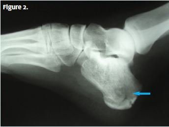
An x-ray is obtained (Figure 2), and it shows a comminuted fracture (arrow) of the calcaneus.
Learnings
Calcaneus fractures account for 1.2% of all fractures in U.S. adults, and they occur most commonly in those who are about 40 years of age. Men are three times more likely than women to sustain such fractures. Most injuries (71%) occur from a fall from a height, usually over 6 feet (1.8 m). Fractures may be intraarticular (75%), which means that they involve the subtalar joint (more severe fractures with worse outcomes), or extra-articular (25%), which means that they do not involve the subtalar joint (and often have a favorable outcome).
What to Look For
During the medical history, check for the following items.
- Onset—gradual versus sudden: Most mechanisms will be a fall from height with sudden onset of pain.
- Location: These fractures are typically over the heel, but there may be referred pain, so even when there is a known mechanism of ankle strain, palpate the heel.
- Duration: Typically patients with these fractures seek immediate medical care, though if there are extenuating circumstances, such as substance use, assault, or physical abuse, the patient may delay seeking care.
- Severity: Pain is typically severe and increases with attempts to bear weight.
- Other types of trauma: Is there ankle, leg, or hip pain? Is there intra-abdominal, chest, neck, or head pain?
- Social history: Ask about the patient’s occupation, ask whether there is alcohol or substance use, and consider assault as a cause.
The following diagnostic tests are appropriate.
- X-rays:
- Obtain lateral and axial views, and consider an oblique view if an avulsion fracture is suspected.
- X-rays are usually adequate for determining the severity of the deformity and assessing the prognosis.
- Computed tomography scans:
- These are useful for fracture evaluation when findings are normal on plain x-rays.
- These are helpful for defining the extent of the fracture to determine surgical indications and approach.
- Magnetic resonance imaging:
- Use this modality to evaluate for stress fracture.
- Use this modality to further define nonspecific computed tomography findings.
The following are indications for transferring the patient to an emergency department:
- An open fracture
- Severe pain
- Possibility of a compartment syndrome
- The presence of neurovascular compromise
- Fractures with dislocation
- Comorbid conditions such as coagulopathy, anticoagulant use, immunosuppression, and difficulty with baseline ambulation