Published on
Differential Diagnosis
- Mallet finger
- Dislocation
- Distal phalanx fracture
- Osteomyelitis
- Osteosarcoma
Diagnosis
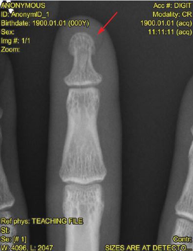
The patient has a nondisplaced distal phalangeal fracture. The most telling sign is the subtle vertical lucency within the distal phalanx (see Figure 2).
Learnings
- A minimum of three x-ray views should be obtained; often, as in this case, the fracture will be seen on only one projection
- Consider localized images (such as a dedicated finger x-ray instead of a hand x-ray) to allow for better resolution and magnification
- Evaluate the x-ray for fracture fragments, lucency, disruption of the trabeculations, or a break in the cortex
- Neurovascular status should be established upon initial assessment
Pearls for Initial Management and Considerations for Transfer
- With evidence of an isolated fracture, splinting and outpatient follow-up is appropriate
- Specifically examine and document presence or absence of associated laceration, as this designates and “open fracture.” This may not change the urgent care management, but should prompt good cleansing as well as aftercare instructions, including warning of signs of infection and the importance of timely follow-up
- If there is an associated subungual hematoma, consider trephination for drainage. Nail removal is rarely required, even with large subungual hematomas
- Transfer with signs of infection, neurovascular compromise, intractable pain, or diagnostic uncertainty
Clinical Challenge: October 2017
1 2