Published on
Differential Diagnosis
- Clavicular fracture
- Shoulder strain
- Fracture of the humerus
- Dislocation of the humerus
- Pneumothorax
Physical Examination
On physical examination, his vital signs are as follows: temperature, 98.6°F (37°C); pulse rate, 112 beats/min; respiration rate, 24 breaths/min; blood pressure, 138/92 mm Hg; and oxygen saturation, 94% on room air. He is alert and oriented, is not in acute distress, and is breathing comfortably. He has no pain on palpation of the posterior cervical spine. He does have pain on palpation at the acromioclavicular joint and minimal swelling at the site, but there are no cuts or breaks in the skin.
Diagnosis
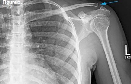
A chest x-ray is obtained (Figure 2) that shows an acromioclavicular joint injury. Note that the distal clavicle and acromion are not approximated, and there is approximately 50% vertical displacement, which means that this is a Rockwood type 3 injury. Rockwood classification is as follows:
- Type 1: The acromioclavicular joint is intact. Treatment can usually be done on an outpatient basis and is conservative.
- Type 2: There is slight vertical separation of the acromioclavicular joint. Treatment is conservative.
- Type 3: The acromioclavicular ligament is disrupted and the acromioclavicular joint is dislocated. The coracoclavicular distance for the injured joint is 25% to 100% greater than for the uninjured joint. Treatment for these injuries is controversial, but they are usually treated conservative initially.
- Type 4: The acromioclavicular joint is dislocated, the coracoclavicular ligaments are completely torn, the clavicle is displaced into the trapezius, and the deltoid and trapezius are detached from the distal clavicle. Treatment for these injuries is surgical repair, and a lidocaine injection may decrease pain.
- Type 5: The coracoclavicular distance for the injured joint is 100% to 300% greater than for the uninjured joint. Treatment is the same as for type 4.
- Type 6: The acromioclavicular joint is dislocated, and the clavicle is in the subcoracoid position. Treatment is the same as for type 4.
Learnings
An acromioclavicular separation is typically caused by a traumatic fall onto the affected shoulder, such as during sports or motor vehicle accidents. The bony approximations superior to the humerus are the distal clavicle and acromion, and the medial approximation is the glenoid cavity. The acromion and clavicle are held together by strong ligaments, which may be stretched or torn with injury. The acromioclavicular ligament provides horizontal stability, whereas the coracoclavicular ligaments provide vertical stability. As with all shoulder injuries, adjacent structures may be damaged. A fracture may occur at the proximal humerus or glenoid cavity.
- The axillary nerve: This nerve runs below the humeral head and is the most commonly injured nerve in shoulder dislocations. It innervates the deltoid and teres minor muscles and skin over the lateral shoulder. Assess its function by checking sensation over the deltoid muscle.
- The axillary artery: Confirm the presence of the distal pulses.
What to Look For
When performing the physical examination, look for the following.
- Mechanism of injury:
- Injuries are common in male intercollegiate athletes, with those participating in rugby, wrestling, and hockey having the highest incidence.
- If the mechanism was a fall, there may be other injuries. Ask about pain in the elbow, wrist, hand, head, and neck.
- Differentiate a mechanical fall from a syncopal episode or a seizure, which may result in other injuries.
- If there was an altercation, ask about other injuries, police reports, and the possibility of physical abuse.
- Neurovascular compromise—ask about numbness or disproportionate pain
- Location and exacerbation of pain:
- Localize the area of greatest pain.
- Ask what makes the pain worse, such as movement, breathing, and palpation.
- Appearance:
- Look for a visible deformity; compare the injured area to the other acromioclavicular joint.
- Assess the integrity of the skin and swelling. Check for fracture, ecchymosis, and erythema.
- Look for an empty sulcus sign, which is a dimpling of the skin just below the acromion seen when looking at the lateral aspect of the shoulder. It may indicate a shoulder dislocation.
- Location of pain—palpate over the acromioclavicular joint to see if this elicits pain
- Range of motion:
- Assess abduction and adduction.
- Assess internal and external rotation.
- Neurovascular status:
- Check pulses (for injury to the axillary artery).
- Assess sensation (for injury to the axillary nerve).
- The one-finger test: Findings on this test are positive when the practitioner asks the patient to point to the area of greatest pain and they point directly to the acromioclavicular joint.
X-ray views to be obtained are the anteroposterior view, the lateral view, and the axillary view, the latter of which is needed to diagnose Rockwood type 4 injuries. Stress views are no longer used. If there is no history of a fall, no swelling, and no pain at rest, and if there is a normal range of motion, the therapeutic utility of x-rays is extremely low.
Indications for transfer to an emergency department include intractable pain, the potential for associated life-threatening injuries, hemodynamic instability, and the possibility of open fracture, infection, pneumothorax, or other sequelae of major trauma.