Published on
Differential Diagnosis
- Acute calcific periarthritis
- Gout
- Infectious arthritis
- Scaphoid fracture
Diagnosis
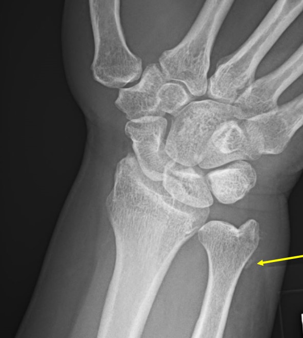
The x-ray shows linear calcification alongside the distal ulna. This patient was diagnosed with acute calcific periarthritis, an acutely painful monoarticular condition where there is juxta-articular deposition of calcium hydroxyapatite crystals and local inflammation.
Learnings/What to Look for
- Acute calcific periarthritis occurs more frequently in females than males, between 40 and 70 years of age
- This condition is a clinical subset of hydroxyapatite deposition disease and occurs when crystals are acutely deposited in the periarticular capsular structures:
- Deposits in tendons result in calcific tendonitis
- Deposits in bursa result in calcific bursitis
- Deposits in the shoulder joint result in Milwaukee shoulder
- Well-circumscribed ovoid or curvilinear calcification may be observed adjacent to a joint (usually on one side)
Pearls for Urgent Care Management
- Acute calcific periarthritis is managed conservatively with NSAIDs but may require corticosteroid injection
- Acute symptom resolution may be achieved within a week
- Calcification significantly decreases in 3-4 weeks, and clears completely in 6-8 weeks
Acknowledgement: Image and case presented by Experity Teleradiology (www.experity.com/teleradiology).
Read More
- A 41-Year-Old With Dorsal Wrist Pain After A Slip-And-Fall
- A 47-Year-Old Male With Chronic Wrist Pain And No Recent Trauma
A 55-Year-Old Female with Sudden-Onset Wrist Pain
1 2