Published on
Differential Diagnosis
- Fibrous dysplasia
- Enchondroma
- Lytic diaphyseal lesion
- Osteomyelitis
Diagnosis
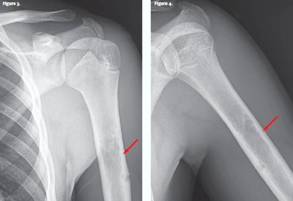
The x-ray reveals an area of mottled lucency mid-diaphysis with endosteal scalloping and cortical breakthrough of the lateral humeral margin. This area corresponded to the site of the patient’s pain. Ill-defined margins were noted (wide zone of transition).
This patient was diagnosed with lytic diaphyseal lesion of the humerus with some aggressive features.
Learnings/What to Look for
- Bony lesions such as osteochondromas and bone cysts can be lead-points for pathologic fractures or injury
- X-Ray is often sufficient to differentiate different bony lesions, but sometimes further imaging studies are required. On a simple radiograph, an ill-defined border with a broad zone of transition is a sign of aggressive growth that is suggestive of either osteomyelitis, eosinophilic granuloma, or a malignant bone tumor
Pearls for Urgent Care Management
- The acute management for pain/injury to bony lesions is similar to fracture management of the same sites, in this case requiring immobilization with a sling similar to a proximal humerus fracture
- It’s critical for the urgent care provider to refer these cases for further evaluation to differentiate a benign lesion from either an infection or malignancy that requires acute management. If the patient is having systemic features (eg, fever, weight loss, etc.), they should be referred immediately to the ED. Without systemic features, the patient can follow-up with an orthopedic specialist as an outpatient.
Acknowledgment: Images and case presented by Experity Teleradiology (www.experity.com/teleradiology).
A 16-Year-Old Boy with Arm Pain After a Baseball Game
1 2