Published on
Differential Diagnosis
- Heterotopic ossification
- Osteosarcoma
- Multiple osteochondromas
- Osteomyelitis
Diagnosis
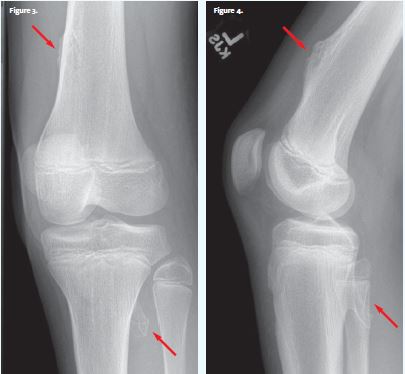
The images reveal irregular prominence of the distal femoral cortex and downward angulated osteophyte at lateral margin proximal tibia and posterior fibula. This patient was diagnosed with multiple osteochondromas.
Learnings/What to Look for
- Osteochondromas are cartilage-capped bony spurs on the external surface of a bone. They grow throughout childhood (while growth plates are open) and the distal femur is the most common location
- Multiple osteochondromas suggests hereditary multiple exostoses (HME), which follows an autosomal dominant inheritance pattern
- Osteochondromas are typically asymptomatic and incidental. They can be palpable and cause pain if associated with local trauma. An osteochroma can be a lead point for a pathologic fracture
- Larger exostoses may impinge on nerves, tendons, or blood vessels, causing extreme pain
- Rarely, osteochondromas can have malignant transformation into a chondrosarcoma
Pearls for Urgent Care Management
- Small exostoses are often incidental, cause no symptoms, and require no treatment, although they can be lead points for pathologic fractures
- Differentiating these exostoses from osteosarcomas and other lytic bony lesions is most important
- Follow-up with a bone specialist is recommended to guide whether monitoring is necessary or to discuss excision if the exostoses are causing impingement symptoms
Acknowledgment: Images and case presented by Experity Teleradiology (www.experityhealth.com/teleradiology).
A 12-Year-Old Boy with Knee Pain After a Baseball Game
1 2