
Published on
Download the article PDF: Ramsay Hunt Syndrome Early Recognition Of Herpes Zoster Oticus In Urgent Care A Case Report
Urgent Message: If not recognized early, herpes zoster oticus may progress to Ramsay Hunt syndrome with facial nerve paralysis. Early antiviral therapy should be initiated, ideally within 72 hours of rash onset, to optimize outcomes.
Vishal Prajapati, MBBS
Keywords: herpes zoster oticus; Ramsay Hunt syndrome; facial nerve palsy; varicella-zoster virus; otalgia; vesicular rash; antiviral therapy
Abstract
Introduction: Herpes zoster oticus results from reactivation of varicella-zoster virus within the geniculate ganglion of the facial nerve. When facial nerve paralysis develops, the condition progresses to Ramsay Hunt syndrome. Early presentations of herpes zoster oticus frequently mimic benign otologic conditions such as otitis externa, creating significant diagnostic challenges in first-contact medical settings. Because vesicular lesions may be sparse at initial presentation, the diagnosis may be easily missed. Prompt recognition and antiviral therapy within 72 hours of rash onset are critical to reducing neural injury.
Presentation: A previously healthy 27-year-old man presented with a 3-day history of severe right-sided burning otalgia, dermatomal hypersensitivity of the auricle and lateral face, and mild serous ear drainage disrupting sleep. He denied fever, vertigo, or hearing loss.
Physical Exam: Inspection of the right auricle revealed translucent vesicles along the conchal bowl and cartilaginous canal with areas of crusting and erythema from ruptured lesions. Mild ear canal edema was present; the tympanic membrane was intact without middle ear effusion. Cranial nerve examination demonstrated normal facial symmetry with full eyelid closure and preserved hearing on bedside testing.
Diagnosis: Early herpes zoster oticus was suspected based on the dermatomal pain pattern and vesicular eruption. Antiviral therapy with valacyclovir was initiated; corticosteroids were deferred given intact facial nerve function. The patient returned 48 hours later with acute lower motor neuron facial palsy (incomplete eyelid closure and drooping of the oral commissure), consistent with a diagnosis of Ramsay Hunt syndrome. Corticosteroid therapy was added, ocular protection measures initiated, and urgent otolaryngology and neurology referrals arranged.
Conclusion: This case highlights that normal cranial nerve findings at initial presentation do not exclude impending facial nerve involvement in herpes zoster oticus. Structured short-interval follow-up within 24–48 hours is recommended to detect potential neurologic progression and initiate timely corticosteroid therapy before permanent facial nerve injury occurs.
Introduction
Acute otalgia is often benign, but a small subset of cases may include early manifestations of neurologic disease requiring prompt recognition and treatment. Herpes zoster oticus results from reactivation of latent varicella-zoster virus (VZV) within the geniculate ganglion of the facial nerve, and viral replication produces inflammation affecting both sensory and motor fibers of the facial nerve.[1] For purposes of this case report, herpes zoster oticus refers to VZV reactivation involving the ear without facial paralysis; when facial nerve paralysis develops in association with auricular vesicles, the condition is referred to as Ramsay Hunt syndrome (RHS).[2]
Patients often present with severe unilateral otalgia, dermatomal hypersensitivity, and subtle vesicular lesions involving the auricle or external auditory canal.[3] Because early dermatologic findings may be sparse or overlooked, herpes zoster oticus may be misdiagnosed as otitis externa, dermatitis, or nonspecific ear pain.3 Early antiviral therapy is most effective when initiated within the first 72 hours of rash onset and may reduce the severity of neurologic complications.[4]
Case Presentation
A 27-year-old previously healthy man presented to a community urgent care clinic with a 3-day history of progressively worsening right-sided ear pain. He described the pain as sharp and burning, radiating across the auricle and lateral face. The discomfort had intensified overnight and was severe enough to disrupt his sleep. The patient reported marked hypersensitivity over the ear and adjacent facial skin.
He denied fever, vertigo, hearing loss, or upper respiratory symptoms. Over the preceding 24 hours, he had noted mild serous drainage from the right ear.
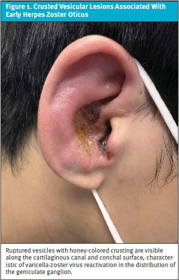
On examination, the patient appeared uncomfortable but was afebrile with normal vital signs. Inspection of the right auricle and external auditory canal revealed several small translucent vesicles along the conchal bowl and cartilaginous canal. Some lesions had ruptured, leaving areas of crusting and erythema (Figure 1). Mild canal edema was present; however, the tympanic membrane appeared intact and without middle ear effusion.
Cranial nerve examination demonstrated normal facial symmetry. The patient could elevate both eyebrows, close both eyes fully, and produce a symmetric smile. His hearing appeared intact on bedside testing.
Medical Decision Making
The primary diagnostic challenge was distinguishing early herpes zoster oticus from uncomplicated otitis externa. The severe neuropathic character of the patient’s pain combined with vesicular lesions suggested viral neuritis rather than simple bacterial infection.
Given the dermatomal pain pattern and vesicular eruption, a clinical diagnosis of early herpes zoster oticus was suspected. Antiviral therapy with valacyclovir 1,000 mg orally 3 times daily for 7 days was initiated. As portions of the canal demonstrated crusting suggestive of secondary bacterial infection, treatment for possible otitis externa was also prescribed.
Early antiviral therapy was initiated to suppress VZV replication and limit inflammatory injury to the facial nerve. As the patient initially had normal facial nerve function, corticosteroids were deferred at the index visit. Instead, the patient was provided explicit counseling regarding symptoms of facial nerve dysfunction and instructed to return promptly if such symptoms occurred.
Differential Diagnosis and Final Diagnosis
Differential diagnoses considered during the initial evaluation included:
- Otitis externa—Common cause of otalgia with canal erythema and discharge.
- Acute otitis media—Less likely given absence of middle-ear findings.
- Auricular dermatitis or eczema—Can produce crusting but usually lacks severe neuropathic pain.
- Herpes zoster oticus—Supported by vesicular lesions and dermatomal neuralgia.
Follow-up and Disposition
The patient returned 2 days later reporting new right-sided facial weakness. The examination revealed diminished right forehead movement, incomplete right eyelid closure (lagophthalmos), and drooping of the right oral commissure. These findings were consistent with Ramsay Hunt syndrome. Notably, the vestibulocochlear nerve was not clinically involved at the second visit. Audiometric testing and vestibular assessment were arranged at the otolaryngology referral to evaluate for eighth cranial nerve complications including sensorineural hearing loss and vertigo, which occur in a subset of patients with RHS.[5]
Systemic corticosteroid therapy was initiated with prednisone 60 mg orally daily for 7 days, followed by a 7-day taper, in combination with continued antiviral therapy.[6] Ocular protection measures were implemented, including lubricating eye drops and nighttime eyelid taping to prevent corneal exposure injury. The patient was referred urgently to otolaryngology and neurology for audiometric testing and further evaluation.
For immunocompetent outpatient presentations such as this case, oral combination therapy is appropriate. Urgent referral to the emergency department for intravenous antiviral therapy (acyclovir 10 mg/kg every 8 hours) should be considered for immunocompromised patients, evidence of disseminated zoster, or an inability to tolerate oral medications.[7]
Discussion
Herpes zoster oticus represents a clinically important cause of acute otalgia in urgent care practice. Reactivation involving the geniculate ganglion may initially present with subtle dermatologic findings and disproportionately severe neuropathic pain.2 Importantly, an atypical variant known as zoster sine herpete—in which VZV reactivation occurs without visible vesicular eruption—has been reported in 8%–25% of patients with acute peripheral facial palsy, reinforcing the need for a high index of suspicion even when skin findings are absent.[8]
The pathophysiology of RHS reflects VZV reactivation producing inflammation and neural edema within the narrow fallopian canal of the temporal bone. Viral spread may involve not only the facial nerve but also the adjacent vestibulocochlear nerve, resulting in sensorineural hearing loss, tinnitus, and vertigo in a significant proportion of patients.5 These eighth cranial nerve complications should be specifically assessed at follow-up and at specialty evaluation. Without treatment, full facial nerve recovery occurs in as few as 20% of patients; this rate improves substantially with combined antiviral and corticosteroid therapy if initiated within 72 hours of onset.6
Corticosteroid dosing for RHS may follow these protocols: prednisone 60 mg orally daily (or 1 mg/kg/day up to 60 mg) for 7–14 days, followed by a gradual taper over an additional 7 days, administered in combination with a 7–10-day course of oral antiviral therapy.6 Corticosteroids may be introduced once facial paralysis becomes apparent to reduce inflammatory edema, and combined therapy may be superior to antivirals alone.6 For immunocompromised patients or those with severe, rapidly progressive palsy, referral for intravenous acyclovir (10 mg/kg every 8 hours for 7 days) is recommended.7
For urgent care clinicians, several diagnostic principles emerge from this case. Severe burning ear pain disproportionate to otoscopic findings should raise suspicion for viral neuritis. A normal cranial nerve examination at the initial visit does not exclude impending facial nerve involvement. Structured short-interval follow-up within 24–48 hours is essential to detect neurological progression and initiate corticosteroid therapy promptly.2 A key clinical pitfall is assuming that severe unilateral otalgia with minimal otoscopic findings represents benign otitis externa; delayed recognition can allow progression to permanent facial nerve injury.
Patients with suspected herpes zoster oticus should receive explicit counseling regarding early symptoms of facial nerve dysfunction, including difficulty closing the eye, asymmetric smiling, or fluid leakage from the mouth, and should be instructed to return immediately if these develop. Because these symptoms often emerge outside the clinical environment, structured follow-up within 24–48 hours may facilitate early recognition of neurological progression and allow timely initiation of corticosteroid therapy.
Clinical Red Flags for Herpes Zoster Oticus in Urgent Care
- Severe unilateral otalgia disproportionate to exam findings
- Burning or neuropathic quality of pain
- Dermatomal hypersensitivity of the auricle or lateral face
- Vesicular lesions within the conchal bowl or external auditory canal
- Hyperacusis (sensitivity to sound) or altered taste
- Early facial heaviness, asymmetry, or difficulty closing the eye
- New vertigo or unilateral hearing loss (suggest eighth cranial nerve involvement)
Ethics Statement
The patient provided written consent for publication of this case report, including the clinical image.
Takeaway Points
- Severe unilateral otalgia with neuropathic features is a hallmark of early herpes zoster oticus and should prompt examination for vesicles and cranial nerve involvement.2
- Early antiviral therapy should be initiated, ideally within 72 hours of rash onset, to optimize outcomes.4
- Antivirals are started at onset of vesicles and corticosteroids (prednisone 60 mg/day for 7–14 days, then tapered) may be added if facial palsy develops.6
- Immunocompromised patients and patients with disseminated zoster warrant emergency referral for intravenous antiviral therapy.7
- Sensorineural hearing loss and vertigo may accompany facial palsy in RHS and require audiometric and vestibular assessment.5
- Follow-up within 24–48 hours is recommended to detect potential progression from herpes zoster oticus to Ramsay Hunt syndrome in order to initiate timely corticosteroid therapy.2
Manuscript submitted March 10, 2026; accepted July 20, 2026.
References
- [1]. Saguil A, Kane S, Mercado M, Lauters R. Herpes zoster and postherpetic neuralgia: prevention and management. Am Fam Physician. 2017;96(10):656-663.
- [2]. Goswami Y, Gaurkar SS. Ramsay Hunt syndrome: an introduction, signs and symptoms, and treatment. Cureus. 2023;15(1):e33688. doi:10.7759/cureus.33688
- [3]. Cunniffe HA, Cunniffe NG. Herpes zoster oticus with meningitis masquerading as malignant otitis externa. BMJ Case Rep. 2019;12(6):e229569. doi:10.1136/bcr-2019-229569
- [4]. Kanerva M, Jones S, Pitkaranta A. Ramsay Hunt syndrome: characteristics and patient self-assessed long-term facial palsy outcome. Eur Arch Otorhinolaryngol. 2020;277(4):1235-1245. doi:10.1007/s00405-020-05817-y
- [5]. Nagel MA, Gilden D. Neurological complications of varicella zoster virus reactivation. Curr Opin Neurol. 2014;27(3):356-360. doi:10.1097/WCO.0000000000000092
- [6]. Monsanto RD, Bittencourt AG, Bobato Neto NJ, Beilke SC, Lorenzetti FT, Salomone R. Treatment and prognosis of facial palsy on Ramsay Hunt syndrome: results based on a review of the literature. Int Arch Otorhinolaryngol. 2016;20(4):394-400. doi:10.1055/s-0036-1584267
- [7]. Uscategui T, Doree C, Chamberlain IJ, Burton MJ. Antiviral therapy for Ramsay Hunt syndrome (herpes zoster oticus with facial palsy) in adults. Cochrane Database Syst Rev. 2008;(4):CD006851. doi:10.1002/14651858.CD006851.pub2.
- [8]. Zhou J, Li J, Ma L, Cao S. Zoster sine herpete: a review. Korean J Pain. 2020;33(3):208-215. doi:10.3344/kjp.2020.33.3.208
Author Affiliation: Vishal Prajapati, MBBS, UCC Urgent Care Centre, Toronto, Ontario, Canada. Author has no relevant financial interests with any ineligible companies.