Published on
The Resolution
Differential Diagnosis
- Sinus tachycardia
- Supraventricular tachycardia
- Atrial fibrillation
- Multifocal atrial tachycardia
- Inferior STEMI
Diagnosis
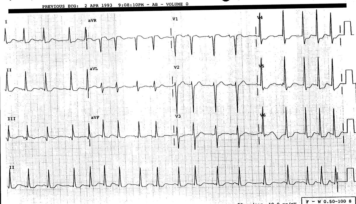
The ECG revels an irregular rhythm, but it is sinus. Notice the different p wave morphology. There are some nonspecific ST changes, but no ST elevation concerning for an acute myocardial infarction. The r waves are prominent consistent with left ventricular hypertrophy (LVH). Q waves are present inferiorly, possibly indicating a prior MI. Arrows (below) point to the “multifocal” p waves.
Learnings
- MAT is an atrial rhythm, and not ventricular.
- Complexes are narrow and irregular, but p waves are present, which would not be the case with atrial fibrillation.
- The ST segments are decreased in the anterior lateral leads (V4-6), but this is a nonspecific finding. Comparison to past ECGs and correlation with the patient’s symptoms are important.
MAT occurs commonly in patients with COPD, and is likely present in our patient given his history of smoking and the scattered wheezing heard on lung auscultation. It may
A 73-Year-Old Man with a 2-Week History of Palpitations
1 2