Published on
Differential Diagnosis
- Sinus tachycardia
- Atrial fibrillation
- Multifocal atrial tachycardia (MAT)
- Atrial flutter with variable atrioventricular (AV) conduction
- Hyperkalemia
Diagnosis
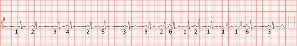
The diagnosis is multifocal atrial tachycardia (MAT). The ECG reveals an irregular, narrow complex rhythm with a ventricular rate of 108 beats per minute. There are at least 3 different P-wave morphologies with variable P-P intervals. This constellation of findings is consistent with multifocal atrial tachycardia. There is also low voltage present, likely secondary to increased impedance from hyperinflated lungs.
MAT is an irregularly irregular rhythm, typically between 100-150 beats per minute, that arises from multiple ectopic foci within the atria. It is most often seen in patients with advanced lung disease, such as COPD. MAT is defined by a rate >100 beats per minute with at least 3 morphologically distinct atrial complexes, varying P-P intervals, and an isoelectric baseline between P waves.1
The mechanism is not completely understood but is believed to be caused by either re-entrant centers, increased atrial automaticity, or triggered centers, and is often associated with respiratory failure. Underlying hypoxia or hypercarbia, right atrial dilation, and increased sympathetic drive are typical physiologic stressors contributing to development of MAT in respiratory failure. Additionally, medications such as beta agonists or theophylline may contribute to development of MAT.1
Clinicians should focus on treatment of the underlying cause. Unfortunately, the development of MAT during an acute illness/exacerbation should be viewed as a poor prognostic indicator, with a significant in-hospital mortality associated during acute illness.1
Other diagnoses to consider with an irregularly irregular rhythm include atrial fibrillation and atrial flutter with variable conduction. With atrial fibrillation, there should be no discernible P waves, and with atrial flutter, “sawtooth” P waves are often visualized (especially in the inferior leads).
What to Look For
- The differential for an irregularly irregular rhythm should focus on atrial fibrillation, atrial flutter with variable atrioventricular block, and MAT.
- MAT is defined by a rate >100 beats per minute with at least 3 morphologically distinct atrial complexes, varying P-P intervals, and an isoelectric baseline between P waves.
Pearls for Management; Considerations for Transfer
- Management should focus on treatment of the underlying cause and not to control the rate.
- Differentiating between other causes of irregularly irregular rhythms is important as therapeutic approaches vary.
- MAT is associated with a poor prognosis when associated with an acute illness. Transfer to a higher level of care should be initiated early.
References
1. Surawicz B, Knilans TK. Chou’s Electrocardiography in Clinical Practice. 6th ed. Elsevier; 2008.