Published on
Differential Diagnosis
- Bony dysplasia
- Chondroid lesion
- Fibrous cortical defect
- Osteoblastoma
- Osteoid osteoma
- Synovial herniation pit of the femoral neck
Diagnosis
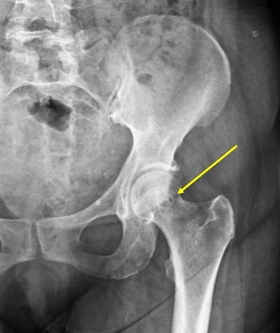
The correct diagnosis is synovial herniation pit of the femoral neck. These are formed by mechanical pressure from the thick anterior hip joint capsule during repetitive hip flexion and extension, which pushes the synovium or soft tissues into the cortical defects in anterior femoral neck. Femoroacetabular impingement may also have a role in their origin. The lesions are acquired and usually stable but can grow over period of time. They could be symptomatic in minority of patients, but typically are incidental findings on the radiographs of asymptomatic patients.
Learnings/What to Look for
- Radiographically, they are visualized as 3 mm to 15 mm diameter round or oval lucent lesions with thin sclerotic margins typically located in anterosuperior femoral neck and 1 cm below the superior neck cortex
- On CT they are low attenuation cortical and subcortical lesions with thin sclerotic margins
- On MRI, the lesion is seen as a smoothly marginated cortical and subcortical mass with low signal on T1 and bright fluid signal on T2 images
- Surrounding bone marrow signal remains normal in bulk of the patients. In symptomatic herniation pit, edema may be present in the surrounding bone marrow
Pearls for Urgent Care Management and Considerations for Transfer
- No therapy is indicated in asymptomatic lesions
- Symptomatic patients with MRI documented bone marrow edema surrounding the herniation pit are treated with intraarticular steroid injection
A 55-Year-Old Female with Hip Pain
1 2