Published on
Differential Diagnosis
- Community acquired pneumonia
- Aspergillus pulmonary infection
- Post-primary tuberculosis
Diagnosis
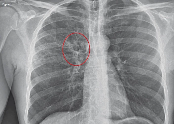
The correct diagnosis in this case is post-primary tuberculosis (TB). This radiograph demonstrates the typical appearance with patchy consolidation and poorly defined linear and nodular opacities. Cavitation is also present. Pleural effusions are also possible but not present in this radiograph.
Post-primary TB is a pattern of disease that arises in a patient who has previously been exposed to TB. This occurs when the disease reactivates in dormant primary lesions, usually several decades after infection when the patient experiences a weakened immune system.
What To Look For
- X-ray findings include interstitial infiltrates, nodular/linear opacities, and/or cavitary lesions commonly in the upper lobes
- Patients with post-primary TB are often asymptomatic or have only minor symptoms, such as a chronic dry cough
- Symptomatic patients experience constitutional symptoms such as fever, malaise, weight loss, or blood-stained productive cough
- Occasionally, patients may present with massive hemoptysis due to an erosion of a bronchial artery
Pearls For Urgent Care Management
- Place the patient in a surgical mask to avoid the spread of TB
- Airborne precautions should be implemented including N95 mask use by healthcare professionals
- If the patient is stable, contact the local public health department to coordinate confirmative testing and a treatment plan
- If the patient is severely ill, transfer to the emergency department for further evaluation and treatment
Image and case provided by Experity Teleradiology (www.experityhealth.com/teleradiology)
Download the article PDF: 51-Year-Old With Hemoptysis
51-Year-Old With Hemoptysis
1 2