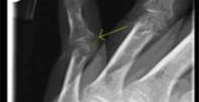
This patient has a volar plate disruption of her R 5th PIP joint.
Initially, subluxation was suspected, a digital block was performed, and the PIP joint was unsuccessfully reduced. Post-reduction films showed mild straightening, but persistent PIP joint hyperextension.
The volar plate forms the floor of the PIP joint, ligamentous at its origin on the proximal phalanx and cartilaginous in its insertion onto the middle phalanx.
A volar plate disruption is usually caused by a hyperextension injury or dislocation of the PIP joint; a mild force may rupture the plate at its distal insertion, causing a swan-neck deformity. Occasionally, a volar plate disruption may also be associated with a fracture of the base of the middle phalanx, which appears on x-ray with a small fragment of bone avulsed from the volar aspect.
As long as the PIP joint is slightly flexed, the joint is considered stable.
A lateral x-ray should be done to confirm that the joint is reduced, and the finger should be splinted in a slightly flexed position with follow-up within three to five days with an orthopedist.
Case presented by Gloria Kim, MD, University Hospitals Urgent Care, Cleveland, OH.
[taq_review]

