Published on
Differential Diagnosis
- Acral lentiginous melanoma
- Atypical nevus
- Talon noir
- Tinea nigra
Diagnosis
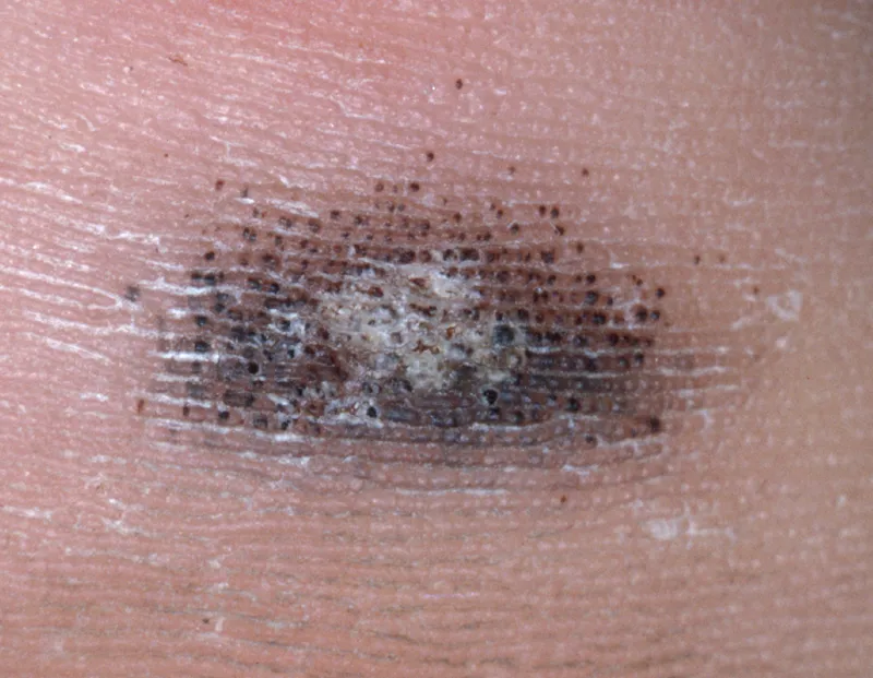
The correct diagnosis is talon noir, also referred to as calcaneal petechiae. Resulting from intraepidermal hemorrhage, this asymptomatic discoloration of acral skin can be caused by shear-force injuries. Talon noir tends to present on the posterior foot, lateral foot, heel, and palm. Lateral shearing forces can cause tearing of blood vessels in the papillary dermis, common in patients who participate in athletic activities. The punctate papillary dermal hemorrhages lead to extravasation of blood into the epidermis and intracorneal retention of hemoglobin. Because of its location in the stratum corneum, it cannot be cleared by phagocytic cells.
What to Look For
- The condition is asymptomatic and painless, so patients may not recall specific etiological events
- On close dermoscopic exam, band like pigmentation may be present
Pearls for Urgent Care Management
- No intervention is required for this condition, it will resolve spontaneously
- Resolution may take 4-6 weeks and may require cessation of triggering activity
- It is important to distinguish from melanoma by its reddish color, sharply defined borders and segmented band like pigmentation
Read More
- Device Aims To Improve Dermatology Referrals
- An Itchy Back With New Moles: A Case Report Of Occult Malignancy
- Talon Noir: A Case Report and Literature Review – PMC
Click Here to download the article PDF.
21-Year-Old With Heel Lesion
1 2