
Published on
Original authors: Ebrahim Barkoudah and Samantha Debold-Hawley
Originally published in 2014. Substantially updated and clinically reviewed in 2026 to reflect current evidence, guidelines, and urgent care practice.
INTRODUCTION
Palpitations are an unpleasant awareness of the heart beating, often described as a sensation of bounding, racing, fluttering, or skipped heartbeats. They are a common reason for urgent care visits. In most cases, palpitations do not imply a pathologic arrhythmia.
The urgent care task is to identify patients whose palpitations may reflect a dangerous arrhythmia or another serious condition, stabilize when necessary, and decide who can be evaluated as an outpatient. History, vital signs, physical examination, and a 12-lead ECG provide the core evaluation. Additional testing should be targeted to the suspected cause rather than ordered routinely.
TRIAGE PEARLS
Check the ABCs (airway, breathing, and circulation).
- Immediate EMS/ED transfer is appropriate when palpitations are accompanied by hypotension, acutely altered mental status, signs of shock, ischemic chest discomfort, acute heart failure, sustained wide-complex tachycardia, syncope during exertion, or another unstable rhythm.
- A heart rate alone does not define instability. A patient with a rate greater than 100 beats/minute may have a physiologic sinus tachycardia, while a slower rhythm may still be dangerous if associated with poor perfusion.
HISTORY PEARLS
- Confirm what the patient means by “palpitations.” Ask whether the sensation is rapid, slow, irregular, pounding, fluttering, or a single skipped beat.
- Determine onset and offset, duration, frequency, regularity, precipitating activity, positional relationship, and what terminates the episode. Having the patient tap out the rhythm can be helpful.
- Syncope or near-syncope with palpitations raises concern for a clinically important arrhythmia, especially when it occurs with exercise or without a prodrome.
- Ask about chest discomfort, dyspnea, diaphoresis, exercise intolerance, neurologic symptoms, fever, blood loss, and symptoms of hyperthyroidism.
- Review caffeine and energy-drink use, nicotine, alcohol, cocaine, methamphetamine and other stimulants, decongestants, beta-agonists, thyroid medication, and drugs that can prolong the QT interval.
- A personal history of structural heart disease, cardiomyopathy, congenital heart disease, prior myocardial infarction, heart failure, or known arrhythmia increases concern. Ask specifically about a family history of sudden unexplained death, inherited arrhythmia syndrome, or cardiomyopathy.
PHYSICAL EXAM PEARLS
- Record heart rate and rhythm, blood pressure, oxygen saturation, temperature, respiratory rate, and general appearance. Orthostatic vital signs are useful when volume depletion, autonomic dysfunction, or postural symptoms are suspected.
- Examine for signs of heart failure, significant murmurs, pulse deficits, poor perfusion, anemia, infection, hyperthyroidism, stimulant use, or other systemic illness.
- A normal examination does not exclude an intermittent arrhythmia.
TESTING PEARLS
Electrocardiography
- Obtain a 12-lead ECG in patients with new, unexplained, recurrent, or clinically concerning palpitations. A normal ECG between episodes does not exclude an intermittent arrhythmia.
- Look for preexcitation (short PR interval with delta wave), long QT, Brugada pattern, conduction disease, ischemic changes, atrial fibrillation/flutter, ectopy, and other rhythm abnormalities.
Targeted laboratory testing
- Laboratory testing should be guided by the history and ECG. Consider glucose, CBC, electrolytes and magnesium, pregnancy testing, or thyroid studies when clinically indicated. In hyperthyroidism, TSH is typically suppressed; free T4 and/or T3 are elevated.
- Routine BNP/NT-proBNP, digoxin levels, urine testing, or broad chemistry panels are not necessary for otherwise stable palpitations without a specific indication.
- Cardiac troponin is not CLIA-waived and is generally not available in typical urgent care centers operating under a Certificate of Waiver. If ischemia, myocarditis, or another troponin-requiring diagnosis is a meaningful concern, transfer for ED/hospital evaluation rather than delaying care for nonwaived testing.
Ambulatory ECG monitoring
- For stable patients with unexplained recurrent palpitations, ambulatory ECG monitoring should match symptom frequency. A 24- to 48-hour Holter is most useful for daily symptoms; patch or external-loop monitoring over days to weeks is more useful for less frequent events.
DIAGNOSIS PEARLS
Diagnosis is done mainly by exclusion—rule out the worst first.
- Common cardiac causes include premature atrial or ventricular beats, supraventricular tachycardia, atrial fibrillation/flutter, sinus tachycardia, preexcitation syndromes, ventricular arrhythmias, structural heart disease, and device malfunction.
- Common noncardiac causes include stimulants and medications, fever, anemia, hypovolemia, hypoglycemia, electrolyte disturbance, hyperthyroidism, pregnancy/menopause-related autonomic symptoms, anxiety or panic, and other causes of increased sympathetic tone.
Selected ECG rhythms
Atrial fibrillation is characterized by absent discrete P waves and an irregularly irregular ventricular response. New AF requires assessment of hemodynamic stability, rate, precipitating illness, duration of symptoms, and thromboembolic risk.
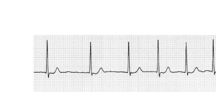
Atrial flutter commonly produces organized flutter waves and may conduct with a fixed or variable ventricular response. It does not inevitably “degenerate” into atrial fibrillation, although the two rhythms can coexist in the same patient.
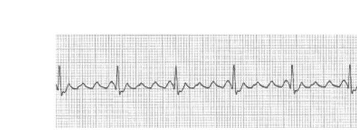
Multifocal atrial tachycardia (MAT) is an irregular narrow-complex tachycardia with at least three P-wave morphologies and a ventricular rate greater than 100/min. When the same atrial pattern occurs at a rate below 100/min, the term wandering atrial pacemaker is preferred. MAT often accompanies pulmonary disease or other acute systemic illness.
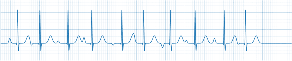
Wolff-Parkinson-White pattern shows ventricular preexcitation, typically a short PR interval and delta wave. The clinically important issue is whether tachyarrhythmia is present and whether atrial fibrillation is preexcited.
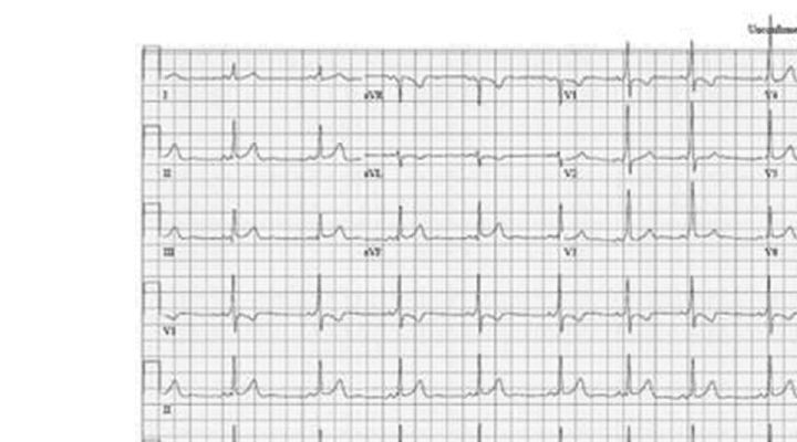
Wide-complex tachycardia should be considered ventricular tachycardia until proven otherwise. A patient can appear relatively stable and still have a potentially life-threatening ventricular arrhythmia.
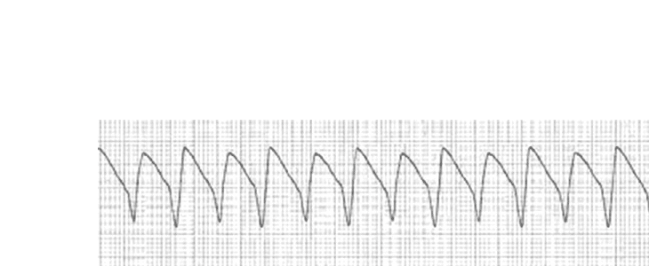
A junctional escape rhythm is generally a narrow-complex rhythm around 40-60 beats/minute with absent, inverted, or retrograde P waves. Management depends on symptoms and perfusion rather than the ECG label alone.

TREATMENT AND DISPOSITION PEARLS
Unstable tachyarrhythmia
- For persistent tachyarrhythmia causing hypotension, acutely altered mental status, shock, ischemic chest discomfort, or acute heart failure, activate EMS/ED transfer immediately. Provide airway support, oxygen if hypoxemic, cardiac monitoring, IV access, and a 12-lead ECG when it does not delay care.
- Synchronized cardioversion is the treatment for unstable tachyarrhythmia with a pulse. Urgent care centers should perform it only when appropriately equipped and staffed; transfer should not be delayed.
Regular narrow-complex SVT
- In a stable patient with regular narrow-complex SVT, attempt a vagal maneuver. A modified Valsalva maneuver is more effective than the traditional maneuver and is preferred when practical.
- Adenosine may be used for a regular narrow-complex tachycardia only in a setting with continuous monitoring, IV access, immediate resuscitation capability, and clinician familiarity. Typical urgent care centers without that capability should arrange EMS/ED transfer.
Wide-complex tachycardia
- Treat a wide-complex tachycardia as ventricular tachycardia until proven otherwise. Adenosine is considered only for a regular monomorphic wide-complex rhythm when the diagnosis remains uncertain and full resuscitation capability is present. Irregular or polymorphic wide-complex rhythms require emergency management.
Atrial fibrillation and flutter
- Transfer patients with AF/flutter who are unstable, have ischemic symptoms, acute heart failure, syncope, stroke symptoms, marked rapid ventricular response, or another serious precipitant. New or first-detected AF generally warrants same-day higher-level evaluation when duration is uncertain or the patient lacks an established management plan.
- For stable known AF with controlled rate, no concerning symptoms, and reliable follow-up, urgent outpatient management may be reasonable depending on the patient’s history and established plan.
- Stroke prevention is based on thromboembolic risk and oral anticoagulation when indicated. Aspirin alone or aspirin plus clopidogrel is not an alternative to anticoagulation for AF stroke prevention.
Preexcitation/WPW
- For regular narrow-complex reentrant SVT in a patient with WPW pattern, vagal maneuvers and adenosine may be used in an appropriately monitored setting.
- Do not give AV-nodal blockers—including adenosine, beta-blockers, diltiazem/verapamil, digoxin, or IV amiodarone—to a patient with preexcited atrial fibrillation. If unstable, perform synchronized cardioversion; stable preexcited AF requires emergency-level management with an appropriate antiarrhythmic such as procainamide or ibutilide.
Sinus tachycardia and ectopy
- Sinus tachycardia is a physiologic response until proven otherwise. Treat the underlying cause—fever, hypovolemia, anemia, pain, hypoxia, stimulant use, hyperthyroidism, or other illness—rather than attempting to “convert” the rhythm with Valsalva.
- Isolated PACs/PVCs in a stable patient with a reassuring ECG and no structural-heart-disease red flags are often benign. Avoid empiric outpatient beta-blocker prescriptions from urgent care unless there is a defined diagnosis and an established treatment plan.
Bradycardia or junctional escape
- Asymptomatic junctional escape related to increased vagal tone may need no acute treatment. Symptomatic bradycardia with hypotension, altered mental status, ischemic discomfort, shock, or acute heart failure requires emergency evaluation; atropine is part of the ACLS bradycardia pathway. Digoxin is not a treatment for junctional escape rhythm.
MEDICOLEGAL PEARLS
- Document vital signs, the 12-lead ECG interpretation, associated chest pain/syncope/dyspnea, relevant stimulant and medication exposure, and personal/family cardiac history.
- Document QT-prolonging medications and electrolyte-risk factors when relevant.
- If pulmonary embolism is a concern, document the clinical PE assessment and use an appropriate PE pathway; a Wells score is not required simply because a patient has palpitations.
- Document the reason for ED transfer or, when outpatient management is chosen, the absence of instability/red flags and the follow-up plan.
References
- Joglar JA, Chung MK, Armbruster AL, et al. 2023 ACC/AHA/ACCP/HRS guideline for the diagnosis and management of atrial fibrillation. Circulation. 2024;149:e1-e156.
- American Heart Association. Adult Tachyarrhythmia With a Pulse Algorithm. 2025.
- Page RL, Joglar JA, Caldwell MA, et al. 2015 ACC/AHA/HRS guideline for the management of adult patients with supraventricular tachycardia. Circulation. 2016;133:e506-e574.
- Al-Khatib SM, Stevenson WG, Ackerman MJ, et al. 2017 AHA/ACC/HRS guideline for management of patients with ventricular arrhythmias and the prevention of sudden cardiac death. Circulation. 2018;138:e272-e391.
- Steinberg JS, Varma N, Cygankiewicz I, et al. 2017 ISHNE-HRS expert consensus statement on ambulatory ECG and external cardiac monitoring/telemetry. Heart Rhythm. 2017;14:e55-e96.
- Appelboam A, Reuben A, Mann C, et al. Postural modification to the standard Valsalva manoeuvre for emergency treatment of supraventricular tachycardias (REVERT): a randomised controlled trial. Lancet. 2015;386:1747-1753.
- Barkoudah E, Collins JP. Management of palpitations in urgent care. J Urgent Care Med. 2012;7:7-12.