Published on
Differential Diagnosis
- Intertrochanteric hip fracture
- Osteosarcoma
- Fracture of the inferior pubic ramus
- Hip osteoarthritis
- Subcapital hip fracture
- Septic arthritis
Physical Examination
On physical examination, her vital signs are as follows: temperature, 99.1°F (37.3°C); pulse rate, 108 beats/min; respiration rate, 20 breaths/min; blood pressure, 164/94 mm Hg; oxygen saturation, 98% on room air. She is alert and oriented, is not in acute distress, and is breathing comfortably.
Externally, her left hip appears normal, without erythema or swelling, and there are no cuts or breaks in the skin. She has moderate pain with flexion and extension of the hip, and with passive internal and external rotation through the range of motion. She has no pain with movement through the range of motion of the left knee. Her neurovascular status is intact, with a 2+ dorsalis pedis pulse, and sensation is grossly intact.
Diagnosis
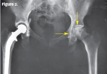
An x-ray (Figure 2) shows arthritic changes (arrows) in the left hip and evidence of replacement of the right hip.
What to Look For
To differentiate between traumatic hip pain and atraumatic pain when obtaining the medical history, first evaluate the mechanism at the onset of pain. Arthritic pain will typically be chronic pain that may have been exacerbated by a specific mechanism. The pain will be worse with movement through the range of motion during weight-bearing. Inquire about ability to ambulate and activities that are limited because of pain, paresthesia, or a sensation of warmth or coolness of the extremity. The past history should include information about any past injuries, surgeries, and therapies.
When performing the physical examination, document the patient’s general appearance and ability to ambulate. Inspect and palpate for skin changes such as erythema, ecchymosis, abrasions, lacerations, fluctuance, necrosis, crepitus, and ecchymosis. Palpate for location of pain, and look for pain exacerbators such as weight-bearing and movement through the range of motion.
A plain x-ray will typically reveal arthritic changes in the hip, but clinical correlation is important. An x-ray of an arthritic hip will show joint-space narrowing, osteophytes, and subchondral sclerosis. The diagnosis requires not only x-ray findings but also symptoms typical of arthritis. Advanced imaging is not required.
Treatment
Symptom management is the initial intervention, through prescribing acetaminophen, exercise, weight loss, icing, movement through the range of motion, and physical therapy. Indications for transfer to an emergency department are as follows:
- Concern that there is a hip fracture
- Intractable pain
- Inability to exclude septic arthritis
Acknowledgment: The image for Figure 1 was produced by Connie Raab as a work of the National Institutes of Health and is in the public domain. Available from: https://commons.wikimedia.org/w/index.php?curid=789996. Figure 2 is an adaptation of Figure 1.