Published on
Physical Examination
On physical examination, the patient’s vital signs are as follows:
- Temperature: afebrile
- Pulse: 98 beats/min
- Respiration: 20 breaths/min
- Blood pressure: 102/74 mm Hg
The patient is alert and oriented. The mid aspect of the right forearm has a bruise over the mid-ulna. There is moderate to severe pain with palpation; she winces with movement of the arm. She has no pain with palpation over the elbow or wrist. She is neurovascularly intact and has a 2+ radial pulse.
Differential Diagnoses
- Smith fracture (distal radius fracture with volar angulation of the fracture fragment)
- Acute elbow dislocation
- Carpometacarpal dislocation
- Colles fracture (distal radius fracture with dorsal angulation of the fracture fragment)
- Acute radius fracture
Diagnosis
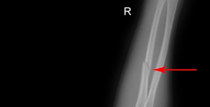
In the radiograph that is obtained (Figure 2), it is obvious that the patient has a nightstick fracture (a midshaft fracture of the ulna).
Learnings
Forearm fractures occur most commonly in men, most commonly from a direct blow to the forearm. A nightstick fracture occurs from a blow to the ulna typically sustained by an arm raised overhead to protect the body.
The two bones making up the forearm are the radius and ulna. These bones rotate to produce supination of the hand (palm facing up) or pronation of the hand (palm facing down). The distal radius juxtaposes against the scaphoid and lunate carpal bones. The carpal bones juxtapose proximally with the distal radius and ulna.
What to Look For
The typical mechanism of injury is an arm raised above the body for protection, with a blow sustained to the mid aspect of the ulna. The medical history should include inquiring about pain proximal and distal to the ulna and looking for a fracture or dislocation in the wrist or elbow. Pain localized only to the mid-ulna is typically from a nightstick fracture. If the injury was sustained in an altercation, there may be other areas of pain that need evaluation.
The physical examination should include documentation of the appearance of the forearm, assessment of the integrity of the skin and swelling, palpation for the area of greatest pain, documentation of the neurovascular status, and palpation as well as determination of the range of motion of the proximal and distal joint. Checking stability before radiology results are available for a patient with an acute injury will be unlikely to change treatment and will likely cause the patient significant pain. Special attention should include evaluation of associated laceration that may be an open fracture or require tetanus prophylaxis.
Treatment
Imaging should include a three-view series of the forearm. Immobilize a nondisplaced fracture in a long arm splint, which may be a sugar tong or ulnar gutter splint. A sling should be given to the patient for comfort. If there is bone shortening or angulation of >10°, the patient should be immediately referred to an emergency department or orthopedist for reduction. Note that unstable fractures include those with >50% displacement, >10° of angulation, involvement of the proximal third of the forearm, or instability at the wrist or elbow.
Pain control should include either hydrocodone-acetaminophen (Vicodin) or oxycodone-acetaminophen (Percocet) and not merely a codeine-containing product. Additional analgesia can be obtained by adding anti-inflammatory medications such as ibuprofen, 600 mg taken three times a day. This can be safely added to the narcotic medication because the nonsteroidal anti-inflammatory drugs are excreted by the kidneys. Other adjunctive therapies include ice and elevation.
If the patient reveals a mechanism of a protective injury, then assessment for other injuries should be done, such as injuries sustained in an assault or from physical abuse.
A nightstick fracture is a mid-ulna fracture typically caused by a defensive posture during an attack. When there is minimal displacement and no shortening, the patient’s arm can be placed in a splint and sling, and the patient can be referred to orthopedics.
Acknowledgment: Image courtesy of Logical Images, Inc. (www.VisualDx.com/JUCM)