Published on
 Differential Diagnosis
- Ankle dislocation
- Jones fracture
- Calcaneal fracture
- Talus fracture
- Bimalleolar fracture
Diagnosis
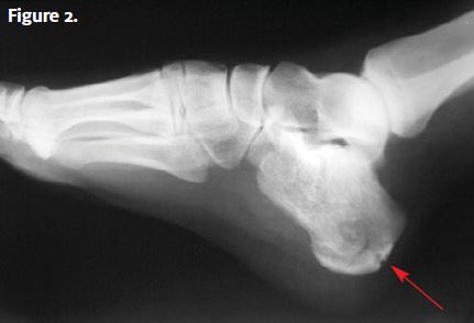
An x-ray (Figure 2) is obtained that shows a comminuted fracture of the calcaneus.
Learnings
Calcaneal fractures account for 1.2% of all fractures in adults, occurring most commonly at the age of 40 years, with the incidence three times higher in men than in women. Most injuries (71%) occur from a fall from a height, usually over 6 feet (0.9 m). When other injuries are present, they are most likely to be to the lower limbs (13%) or the spine (6%). Even with appropriately treated fractures, there is significant morbidity with prolonged pain and disability, which is increased if the fractures are not recognized or appropriately treated. The poor prognosis may result from direct trauma to the articular surfaces, calcaneal fat pad, and peroneal tendons. Fractures are divided into intra-articular (75%), which involve the subtalar joint (more severe fractures having worse outcomes), and extra-articular (25%), which do not involve the subtalar joint (these often have a favorable outcome).
The talus sits on top of the calcaneus, and together these bones make up the hindfoot. The calcaneus approximates distally with cuboid bone. The subtalar joint is the junction of the three facets (anterior, middle, and posterior) between the calcaneus and talus. The posterior aspect of the talus is the tuberosity, the site of the insertion of the Achilles tendon (on the superior aspect).
The calcaneus is the first bone to be impacted with walking or with a fall from a height. Depending on the position of the foot (valgus or varus), the overlying talus bone will transmit a force to a unique portion of the calcaneus, resulting in specific types of fractures.
What to Look For
When performing the physical examination, ask questions about the following.
- Onset: Ask whether the onset was gradual versus sudden. Most often, the mechanism will be a fall from height with sudden onset of pain.
- Location: The typical location is over the heel, but there may be referred pain, so even with a mechanism of ankle strain, palpate the heel.
- Duration: The typical timeline involves the patient seeking immediate medical care, though if there are extenuating circumstances, such as substance use, assault, or abuse, the patient may delay seeking care.
- Severity: Pain is typically severe and increases with any attempt to bear weight.
- Other trauma: Ask about trauma to the ankle, leg or hip pain, intraabdominal pain, or pain in the chest, neck, and head.
- Social history: Inquire about occupation, alcohol or substance use, and any possibility of assault.
Do the following:
- Examine the foot, ankle, knee, and hip.
- Inspection of the foot for signs of swelling, abrasions, and lacerations. Ecchymosis is not a sensitive finding but is specific for a calcaneal fracture.
- Palpate the heel and ankle.
- Assess the range of motion. This evaluation may be deferred when there is severe pain.
- Check the neurovascular status, with documentation of pulses (dorsalis pedis and posterior tibial) and of gross sensation.
- Look for compartment syndrome, which may be a consideration with massive swelling. It is present in 10% of patients with a calcaneal fracture, and it is typically caused by a high-energy deceleration injury.
Keep these important points in mind:
- When a patient has an impact high enough to cause a fracture of the calcaneus, there are often other associated injuries. Perform a complete trauma evaluation, looking specifically for other limb injuries as well as injury to the lower back.
- Document a neurovascular examination. If there is any possibility of compartment syndrome, immediately transfer the patient to an emergency department for orthopedic consultation and measurement of compartment pressures.
Most of these patients will be sent to an emergency department. Indications for transfer include the following:
- Open fracture
- Severe pain
- Possibility of compartment syndrome
- Neurovascular compromise
- Fractures with dislocation
- Possible comorbid conditions such as coagulopathy, anticoagulant use, immunosuppression, and difficulty with baseline ambulation
Acknowledgment: Figure 1 is used with permission under a GNU Free Document License, Version 1.2, from Jojo. Original figure available from https://upload.wikimedia.org/wikipedia/commons/e/e8/Calcaneus_Fracture.jpg.