Adult with Painful Lesion That Enlarges and Darkens

Adult with Painful Lesion That Enlarges and Darkens

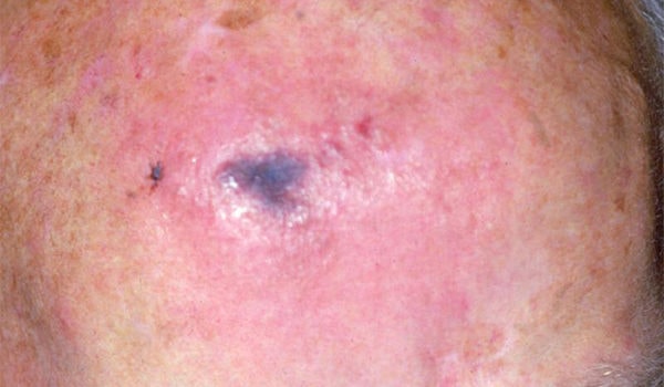
An 80-year-old man reports waking up with a localized area of erythema on his forehead. He says that he did not worry about it until the lesion slowly became white and then started turning blue over a few hours. He says that the lesion is painful and is getting larger and darker. He reports no headaches, muscle pain, abdominal pain, sweating, or tremors. View the image taken (Figure 1) and consider what your diagnosis would …

Read More
Skin Rash with Pustules on a Newborn

Skin Rash with Pustules on a Newborn

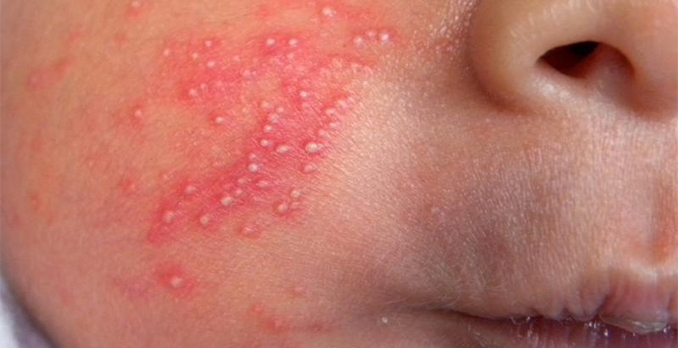
A healthy, full-term baby girl was born to a healthy mother who had adequate prenatal care and no signs of infectious diseases. Three days after birth, the baby developed multiple 1-mm pale-yellow to yellow pustules within a large inflammatory wheal on her cheek. Several hours later, the rash starts to resolve. View the image taken (Figure 1) and consider what your diagnosis would be.

Read More
Eye Redness, Pain, and Light Sensitivity

Eye Redness, Pain, and Light Sensitivity

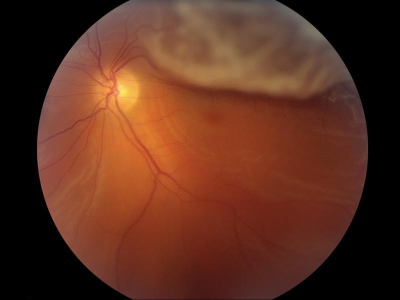
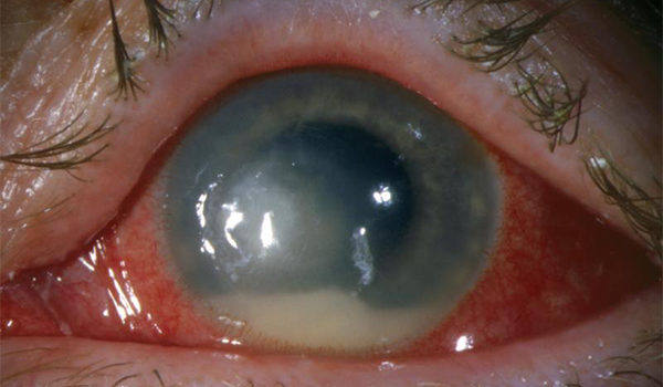
A 30-year-old woman presents with a red eye, associated with eye pain, light sensitivity, and slightly decreased vision, which started 4 days earlier. She was previously treated with tobramycin, but she has not noticed any change in the condition of her eye. Examination shows an injected conjunctiva and a hypopyon. On further examination with a slit lamp, inflammatory cells are evident in the anterior chamber. View the image taken (Figure 1) and consider what your …

Read More
Adult with Eye Pain, Photophobia, and Decreased Vision

Adult with Eye Pain, Photophobia, and Decreased Vision

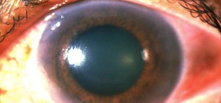
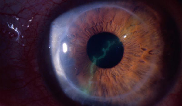
A 35-year-old woman presents with a history of 1 day of pain in her right eye, photophobia, and decreased vision. There is no history of trauma. One day earlier, she was seen at an urgent care center, where the diagnosis was a corneal abrasion, and she was treated with topical trimethoprim-polymyxin. Her symptoms have worsened, so she has returned for further evaluation. View the image taken (Figure 1) and consider what your diagnosis would be.

Read More
Log In