Published on
RESOLUTION
Differential Diagnoses
- Cardiomegaly from heart failure
- Pericardial tamponade
- Esophageal rupture (Boerhaave syndrome)
- Left lower lobe infiltrate
- Pleural effusion
Physical Examination
On examination, the patient is found to be afebrile and to have a pulse of 112 beats/min, a respiration rate of 20 breaths/min, and a blood pressure of 97/54 mm Hg. Alert and oriented, he seems minimally uncomfortable, speaks normally, and provides a good medical history. Both of his lungs are clear to auscultation. He has a regular heart rate and rhythm without murmurs, rub, or gallop. His abdomen is soft and nontender, without rigidity, rebound, or guarding. In the extremities, his peripheral are pulses 2+ and equal.
Diagnosis
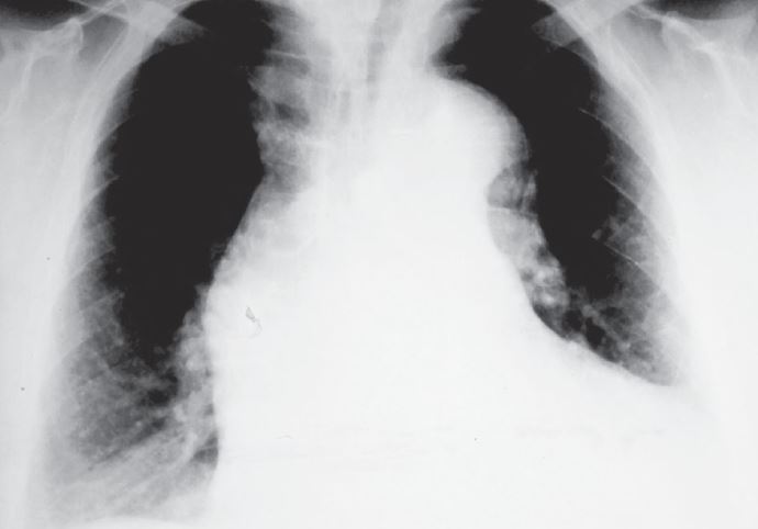
Because of radiographic findings (Figure 2), the diagnosis is acute aortic dissection.
Learnings
Patients with aortic dissection can often present in an innocuous manner, but rapid diagnosis is vitally important, because mortality is as high as 58% without surgery, increasing by 1% to 2% per hour of delayed diagnosis.
Complicating manners, 15% of patients have a painless dissection, and 13% present as having syncope. Risk factors include male sex, hypertension, cocaine use, family history, and connective-tissue diseases such as Marfan syndrome and Ehlers-Danlos syndrome.
The aorta is the main outflow of the heart, taking blood from the left ventricle, through the aortic valve into the aortic arch and its three main branches. Those three branches are (proximal to distal) the brachiocephalic artery, which divides into the right subclavian artery and the right internal carotid artery; the left carotid artery; and the left subclavian artery. If the dissection progresses to involve the coronary arteries, the patient with have symptoms of myocardial infarction, whereas if it involves the pericardial space, the patient will have symptoms of tamponade. Involvement of the carotid arteries may manifest with neurologic symptoms or stroke.
What to Look For
Normal findings on chest x-rays cannot be used to rule out a dissection, but if there are findings, they may include the following:
- Widened mediastinum
- Rightward tracheal displacement
- Irregular aortic contour—loss of aortic knob
- Indistinct aortopulmonary window
- Left pleural effusion
With suspicion of aortic dissection and unstable vital signs, activate the emergency medical system and immediately transfer the patient to an emergency department. Inform the emergency medical system that the patient’s condition is unstable and will require emergency transfer with lights and sirens. If there is time, place two intravenous lines and obtain an electrocardiograph. Chest x-ray findings will not change treatment, so do not have the patient leave the urgent care examination room to walk down the hall and get a chest x-ray. Call the emergency department and speak with the physician who will be caring for the patient to inform them that there is an unstable patient with likely aortic dissection who will arrive shortly. This may help with notification of the vascular surgeon and preparation of an operating room.
Sources of delay in transfer due to delay in diagnosis include patients with a blood pressure of more than 105 mm Hg, fever, female sex, and transfer from a tertiary facility. It is important to diagnose aortic dissection rapidly, and the condition should be suspected with sudden onset of severe chest pain as well as with chest pain associated with headache, numbness, neck pain, or limb ischemia.
Acknowledgment: Image adapted from and reused with permission from J. Heuser under a GNU Free Documentation License, version 1.2 (https://en.wikipedia.org/wiki/GNU_Free_Documentation_License). Original image available from https://commons. wikimedia.org/wiki/File:AoDiss_ChestXRay.jpg.