Published on
Resolution
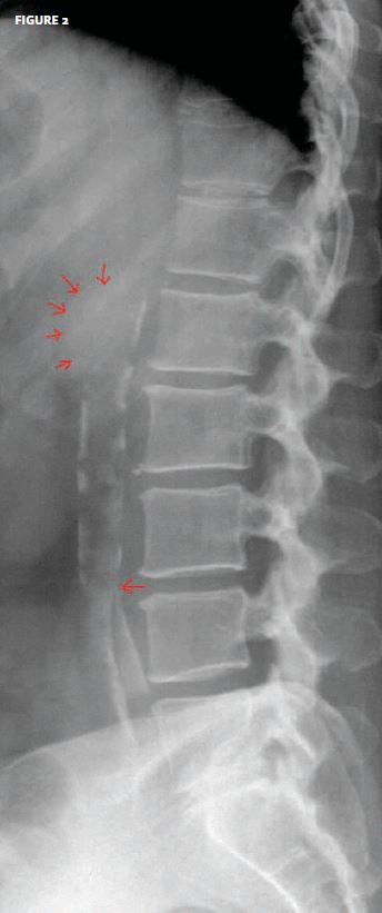
The patient had an aortic aneurysm.
This is quite a dramatic example of calcification of the aorta. At the top end, one sees an aneurismal dilatation of the aorta; at the lower end, one sees the iliac bifurcation.
This patient was referred back to his physician with clear instructions to further evaluate the aneurysm.
Given the patient’s complaint of back pain, additional evaluation with computed axial tomography or ultrasound should be considered.
Acknowledgement: Case presented by Nahum Kovalski, BSc, MDCM, Terem Emergency Medical Centers, Jerusalem, Israel.
These cases are among hundreds that can be found in Terem’s online x-ray Teaching File, with more being added daily. Free access to the file is available at https://www2.teremi.com/xrayteach/. A no-cost, brief registration is required.