This x-ray, taken on a 17-year-old female, represents an incidental finding in her right ankle. View the image taken (Figure 1) and consider what your diagnosis would be.
Read More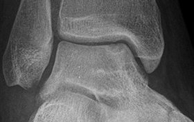

This x-ray, taken on a 17-year-old female, represents an incidental finding in her right ankle. View the image taken (Figure 1) and consider what your diagnosis would be.
Read More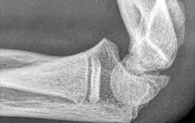
The patient, a 12-year-old boy, suffered a blow to his left elbow. View the image taken (Figure 1) and consider what your diagnosis would be.
Read MoreThe patient, a 4-year-old boy, tripped and twisted his right ankle. He was not able to bear weight on his right foot. View the image taken (Figure 1) and consider what your diagnosis would be. Resolution of the case is described on the next page.
Read More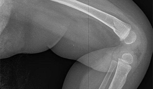
The patient, an 8-month-old boy, was brought in by a caretaker, who reported that the infant had stopped crawling. View the image taken (Figure 1) and consider what your diagnosis would be.
Read More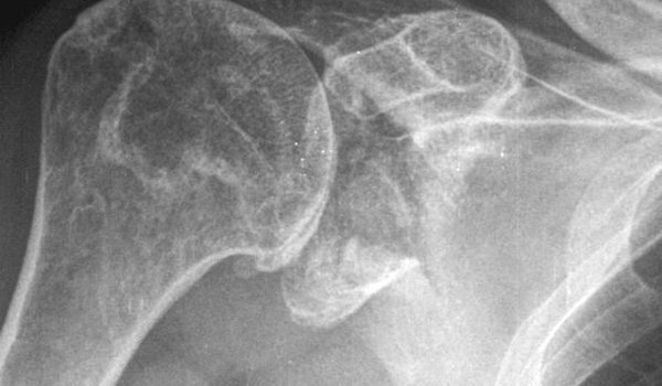
The patient, a 76-year-old male, presented after a blow to his right shoulder. View the image taken (Figure 1) and consider what your diagnosis would be.
Read More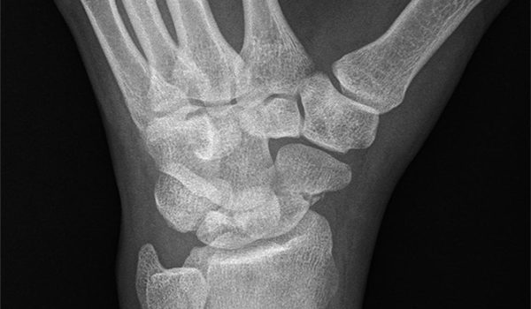
The patient, a 23-year-old male, presented after a blow to his left wrist. View the image taken (Figure 1) and consider what your diagnosis would be.
Read MoreIn each issue, JUCM will challenge your diagnostic acumen with a glimpse of x-rays, electrocardiograms, and photographs of dermatologic conditions that real urgent care patients have presented with. If you would like to submit a case for consideration, please email the relevant materials and presenting information to [email protected]. The patient, a 37-year-old woman, presented after a blow to her left hand. View the image taken (Figure 1) and consider what your diagnosis would be. Resolution …
Read More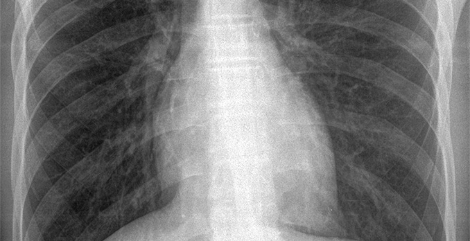
The patient, a 15-year-old male, presented with complaints of chest pain. View the image taken (Figure 1) and consider what your diagnosis would be.
Read More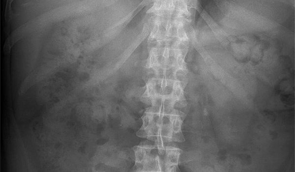
The patient, a 53-year-old woman, presented with nonspecific complaints of significant abdominal pain. View the image taken (Figure 1) and consider what your diagnosis would be.
Read More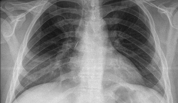
The patient, a 25-year-old female with autism, presented with a 1-week history of fever. View the image taken (Figure 1) and consider what your diagnosis would be. Diagnosis: The x-ray reveals a foreign body in the right airway (arrow) and an infiltrate in the right lower lobe (circle). Referral to a hospital for extraction of the foreign body and treatment is necessary for this patient. Case presented by Nahum Kovalski, BSc, MDCM, Terem Emergency Medical …
Read More