Published on
Differential Diagnosis
- Brugada syndrome
- Hyperkalemia
- Left bundle branch block
- Right bundle branch block
- Wolff-Parkinson-White
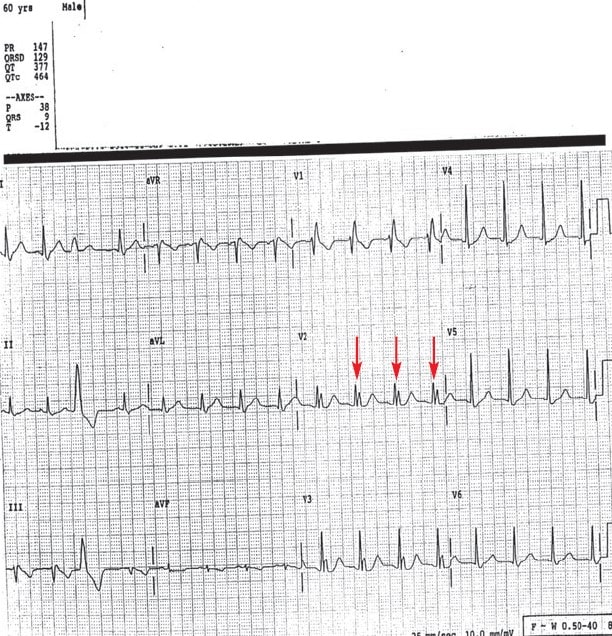
Figure 2.
Diagnosis
This patient has right bundle branch block. The ECG reveals normal sinus rhythm with a prolonged QRS complex (normal is 80-120 ms) and an RSR’ in the right-sided precordial leads (V1-3), consistent with right bundle branch block (RBBB). There is one PVC.
Learnings/what to look for:
- An RBBB occurs as depolarization is delayed when progressing from the left ventricle across the septum to the right ventricle. The initial part of the depolarization is the left ventricle and the subsequent is the right ventricle
- The ECG changes include a widened QRS complex (>120ms), RSR’ in the right precordial leads (V1-3), and a wide and slurred S wave in the lateral leads (I, aVL, V5-6)
- Causes include idiopathic degeneration of the right bundle branch, right ventricular hypotrophy, ischemia, myocarditis, pulmonary embolus, and cardiomyopathy
Pearls for Initial Management and Considerations for Transfer
- Compare with a previous ECG, if available
- If there is concurrent concern for more serious disease such as ischemia or pulmonary embolism, then emergent transfer is indicated
- An incidental finding is common and does not typically require further investigation unless there are concerning clinical features
A 60-Year-Old Man with a 2-Year History of Dizziness
1 2