Published on
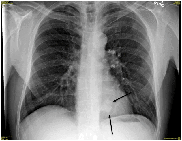
Figure 2.
Diagnosis
The x-ray reveals a left lower lobe lung mass; see the oval-shaped density in the medial aspect of the left lower lobe, visible in the retro cardiac region.
Learnings
- Search pattern on chest x-ray should always include the “hidden areas,” such as the retro cardiac region, lung apices, and hilar regions
- Any increased density in the retro cardiac region or loss of clearly defined left heart border should raise concern for mass or infiltrate
Pearls for Urgent Care Management and Consideration for Transfer
- With this presentation and radiographic findings, the patient needs outpatient referral for advanced imaging and management
A 51-Year-Old Male with a Persistent Cough
1 2